. 2018 Mar 16;8(1):4714.
doi: 10.1038/s41598-018-23142-7.

Non-invasive Estimation of the Intracranial Pressure Waveform from the Central Arterial Blood Pressure Waveform in Idiopathic Normal Pressure Hydrocephalus Patients

Affiliations

Non-invasive Estimation of the Intracranial Pressure Waveform from the Central Arterial Blood Pressure Waveform in Idiopathic Normal Pressure Hydrocephalus Patients

Karen Brastad Evensen et al. Sci Rep. .

Abstract

This study explored the hypothesis that the central aortic blood pressure (BP) waveform may be used for non-invasive estimation of the intracranial pressure (ICP) waveform. Simultaneous invasive ICP and radial artery BP waveforms were measured in 29 individuals with idiopathic normal pressure hydrocephalus (iNPH). The central aortic BP waveforms were estimated from the radial artery BP waveforms using the SphygmoCor system. For each individual, a transfer function estimate between the central aortic BP and the invasive ICP waveforms was found (Intra-patient approach). Thereafter, the transfer function estimate that gave the best fit was chosen and applied to the other individuals (Inter-patient approach). To validate the results, ICP waveform parameters were calculated for the estimates and the measured golden standard. For the Intra-patient approach, the mean absolute difference in invasive versus non-invasive mean ICP wave amplitude was 1.9 ± 1.0 mmHg among the 29 individuals. Correspondingly, the Inter-patient approach resulted in a mean absolute difference of 1.6 ± 1.0 mmHg for the 29 individuals. This method gave a fairly good estimate of the wave for about a third of the individuals, but the variability is quite large. This approach is therefore not a reliable method for use in clinical patient management.


Conflict of interest statement

KBE, FP and SH declare no conflicts of interest. MOR is founding director of AtCor Medical, manufacturer of the SphygmoCor system. PKE has a financial interest in the software company (dPCom AS, Oslo) manufacturing the software (Sensometrics Software) used for analysis of the ICP recordings.

Figures

Figure 1
Figure 1
Non-invasive estimation of ICP waveforms from central aortic BP waveforms. (a) In this study, central aortic BP waveforms were used as input for estimation of non-invasive ICP signals, when the system from the heart to the cranium was said to be unknown. (b) A system description in the frequency domain is found from the transfer function H(f). (c) An estimate for the system is found based on the power spectral density of the central aortic BP waveforms (dotted line) and ICP waveforms (continuous line).
Figure 2
Figure 2
The non-invasive ICP waveform estimated from the Intra-patient approach superimposed on the invasive ICP waveform. For six different time windows of six second duration, the invasive ICP raw signal (continuous red line) is shown superimposed on the estimated non-invasive ICP signal (interrupted blue line) for patient 20. The estimate is established with the Intra-patient approach and shown for the time points (a) 00:00, (b) 1 hour after 00:00, (c) 2 hours after 00:00, (d) 4 hours after 00:00, (e) 7 hours after 00:00, and (f) 9 hours after 00:00. For visual comparison, the estimated ICP signal is time-shifted to match the invasive ICP signal for each time window.
Figure 3
Figure 3
The non-invasive ICP waveform estimated from the Inter-patient approach superimposed on the invasive ICP waveform. For six different time windows of six second duration, the invasive ICP raw signal (continuous red line) superimposed on the estimated non-invasive ICP signal (interrupted blue line) are shown for one patient, established with the Inter-patient approach. The results are shown for the time points (a) 00:00, (b) 1 hour after 00:00, (c) 2 hours after 00:00, (d) 4 hours after 00:00, (e) 7 hours after 00:00, and (f) 9 hours after 00:00. For visual comparison the estimated non-invasive ICP signal is time-shifted to match the invasive ICP signal for each time window.
Figure 4
Figure 4
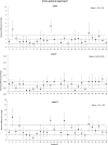
Differences in absolute pulsatile pressure parameters between measured ICP and estimated pulsatile ICP according to Intra-patient approach. The averaged absolute difference between the measured ICP and the estimated ICP is shown for the Intra-patient approach. This is shown for each time domain waveform parameter for each patient ID, with the patient specific standard deviation illustrated as error bars. The total mean for the patient cohort together with its standard deviation is illustrated with dotted lines.
Figure 5
Figure 5
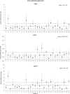
Differences in absolute pulsatile pressure parameters between measured ICP and estimated pulsatile ICP according to Inter-patient approach. The averaged absolute difference between the measured ICP and the estimated ICP is shown for the Inter-patient approach. This is shown for each time domain waveform parameter for each patient ID, with the patient specific standard deviation illustrated as error bars. The total mean for the patient cohort together with its standard deviation is illustrated with the dotted lines.
Figure 6
Figure 6
Association between height and weight with absolute differences in MWA, as estimated from the Intra- and Inter-patient approaches. The associations between the height of the patients and the absolute difference in MWA between measured and estimated ICP waveforms were determined according to (a) the Intra-patient approach and (b) the Inter-patient approach. Further, the association between the weight of the patients and the absolute difference in MWA between measured and estimated ICP waveforms were determined according to (c) the Intra-patient approach and (d) the Inter-patient approach. For each plot the fit line and the Pearson correlation coefficient (R) with significance level is presented.

References

    1. Czosnyka M. Monitoring and interpretation of intracranial pressure. J. Neurol. Neurosurg. Psychiatry. 2004;75:813–821. doi: 10.1136/jnnp.2003.033126. - DOI - PMC - PubMed
    1. Binz DD, Toussaint LG, 3rd, Friedman JA. Hemorrhagic complications of ventriculostomy placement: a meta-analysis. Neurocritical care. 2009;10:253–256. doi: 10.1007/s12028-009-9193-0. - DOI - PubMed
    1. Popovic D, Lee KM, Noninvasive S. monitoring of intracranial pressure. Recent Patents Biomed Engineer. 2009;2:165–179. doi: 10.2174/1874764710902030165. - DOI
    1. Zhang, X. et al. Invasive and noninvasive means of measuring intracranial pressure: a review. Physiol. Meas. (2017). - PubMed
    1. Robba C, et al. Non-invasive assessment of intracranial pressure. Acta Neurol. Scand. 2016;134:4–21. doi: 10.1111/ane.12527. - DOI - PubMed

Publication types