

Production of antibody against elephant endotheliotropic herpesvirus (EEHV) unveils tissue tropisms and routes of viral transmission in EEHV-infected Asian elephants
- PMID: 29549315
- PMCID: PMC5856810
- DOI: 10.1038/s41598-018-22968-5
Production of antibody against elephant endotheliotropic herpesvirus (EEHV) unveils tissue tropisms and routes of viral transmission in EEHV-infected Asian elephants
Abstract
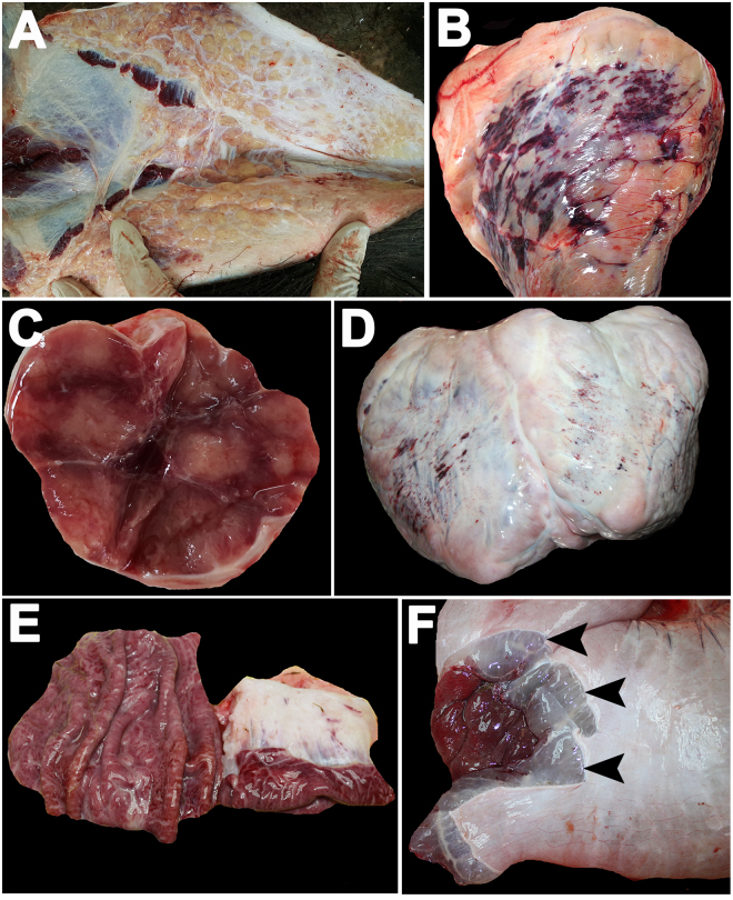
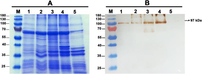
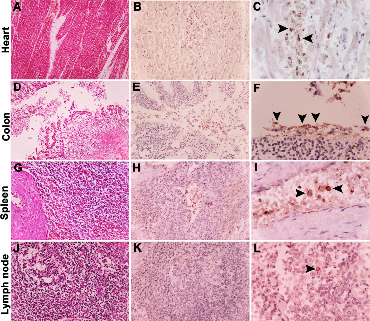
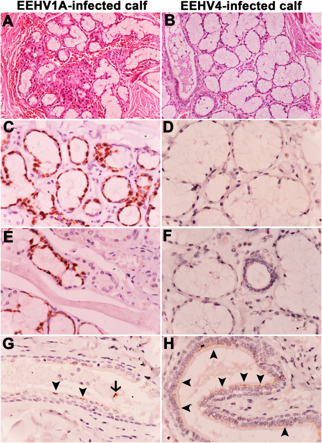
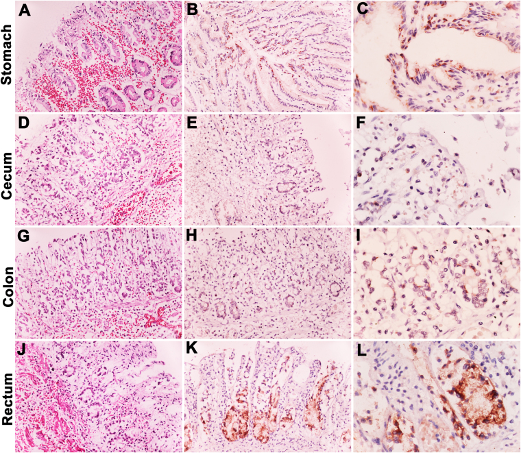
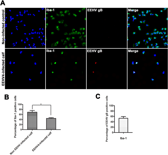
Elephant endotheliotropic herpesvirus (EEHV) is one of the most devastating viral infectious diseases in elephants worldwide. To date, it remains unclear how elephants get infected by the virus, where the virus persists, and what mechanisms drive the pathogenesis of the disease. The present study was aimed to develop an antibody against glycoprotein B (gB) of EEHV, investigate the EEHV tissue tropisms, and provide the possible routes of EEHV transmission in Asian elephants. Samples from elephant organs that had died from EEHV1A and EEHV4 infections, peripheral blood mononuclear cells (PBMC) from EEHV4- and non-EEHV-infected calves were used in this study. The results of western immunoblotting indicated that the antibody can be used for detection of gB antigens in both EEHV1A- and EEHV4-infected samples. Immunohistochemical detection indicated that the EEHV gB antigens were distributed mainly in the epithelial cells of the salivary glands, stomach and intestines. Immunofluorescence test of PBMC for EEHV gB in the EEHV4-infected calf indicated that the virus was observed predominantly in the mononuclear phagocytic cells. The findings in the present study unveil tissue tropisms in the EEHV1A- and EEHV4-infected calves and point out that saliva and intestinal content are likely sources for virus transmission in EEHV-infected Asian elephants.
Conflict of interest statement
The authors declare no competing interests.
Figures
Similar articles
-
Elephant Endotheliotropic Herpesvirus Is Omnipresent in Elephants in European Zoos and an Asian Elephant Range Country.Viruses. 2021 Feb 11;13(2):283. doi: 10.3390/v13020283. Viruses. 2021. PMID: 33670367 Free PMC article.
-
Lethal Hemorrhagic Disease and Clinical Illness Associated with Elephant Endotheliotropic Herpesvirus 1 Are Caused by Primary Infection: Implications for the Detection of Diagnostic Proteins.J Virol. 2020 Jan 17;94(3):e01528-19. doi: 10.1128/JVI.01528-19. Print 2020 Jan 17. J Virol. 2020. PMID: 31723022 Free PMC article.
-
Possible roles of monocytes/macrophages in response to elephant endotheliotropic herpesvirus (EEHV) infections in Asian elephants (Elephas maximus).PLoS One. 2019 Sep 6;14(9):e0222158. doi: 10.1371/journal.pone.0222158. eCollection 2019. PLoS One. 2019. PMID: 31491031 Free PMC article.
-
The occurrence of elephant endotheliotropic herpesvirus in captive Asian elephants (Elephas maximus): first case of EEHV4 in Asia.J Zoo Wildl Med. 2013 Mar;44(1):100-4. doi: 10.1638/1042-7260-44.1.100. J Zoo Wildl Med. 2013. PMID: 23505709
-
Review of Elephant Endotheliotropic Herpesviruses and Acute Hemorrhagic Disease.ILAR J. 2016;56(3):283-96. doi: 10.1093/ilar/ilv041. ILAR J. 2016. PMID: 26912715 Free PMC article. Review.
Cited by
-
Elephant Endotheliotropic Herpesvirus Is Omnipresent in Elephants in European Zoos and an Asian Elephant Range Country.Viruses. 2021 Feb 11;13(2):283. doi: 10.3390/v13020283. Viruses. 2021. PMID: 33670367 Free PMC article.
-
Tissue and cellular tropism of elephant endotheliotropic herpesvirus (EEHV)1A in hemorrhagic disease.PLoS One. 2025 Sep 2;20(9):e0330631. doi: 10.1371/journal.pone.0330631. eCollection 2025. PLoS One. 2025. PMID: 40892760 Free PMC article.
-
Fatal Elephant Endotheliotropic Herpesvirus Infection of Two Young Asian Elephants.Microorganisms. 2019 Sep 26;7(10):396. doi: 10.3390/microorganisms7100396. Microorganisms. 2019. PMID: 31561506 Free PMC article.
-
In vivo characterization of target cells for acute elephant endotheliotropic herpesvirus (EEHV) infection in Asian elephants (Elephas maximus).Sci Rep. 2020 Jul 9;10(1):11402. doi: 10.1038/s41598-020-68413-4. Sci Rep. 2020. PMID: 32647124 Free PMC article.
-
Survival analysis of confirmed elephant endotheliotropic herpes virus cases in Thailand from 2006 - 2018.PLoS One. 2019 Jul 5;14(7):e0219288. doi: 10.1371/journal.pone.0219288. eCollection 2019. PLoS One. 2019. PMID: 31276571 Free PMC article.
References
Publication types
MeSH terms
Substances
LinkOut - more resources
Full Text Sources
Other Literature Sources
