. 2018 Mar 16;8(1):4743.
doi: 10.1038/s41598-018-22853-1.

Prognostic Impact of the Findings on Thin-Section Computed Tomography in stage I lung adenocarcinoma with visceral pleural invasion

Affiliations

Prognostic Impact of the Findings on Thin-Section Computed Tomography in stage I lung adenocarcinoma with visceral pleural invasion

Mei Yuan et al. Sci Rep. .

Abstract

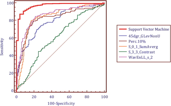
Visceral pleural invasion (VPI) in stageI lung adenocarcinoma is an independent negative prognostic factor. However, no studies proved any morphologic pattern could be referred to as a prognostic factor. Thus, we aim to investigate the potential prognostic impact of VPI by extracting high-dimensional radiomics features on thin-section computed tomography (CT). A total of 327 surgically resected pathological-N0M0 lung adenocarcinoma 3 cm or less in size were evaluated. Radiomics signature was generated by calculating the contribution weight of each feature and validated using repeated leaving-one-out ten-fold cross-validation approach. The accuracy of proposed radiomics signature for predicting VPI achieved 90.5% with ROC analysis (AUC, 0.938, sensitivity, 90.6%, specificity, 93.2%, PPV: 91.2, NPV: 92.8). The cut-off value allowed separation of patients in the validation data into high-risk and low-risk groups with an odds ratio 12.01. Radiomics signature showed a concordance index of 0.895 and AIC value of 88.9% with regression analysis. Among these radiomics features, percentile 10%, wavEnLL_S_2, S_0_1_SumAverage represented as independent factors for determining VPI. Results suggested that radiomics signature on CT exhibited as an independent prognostic factor in discriminating VPI in lung adenocarcinoma and could potentially help to discriminate the prognosis difference in stage I lung adenocarcinoma.


Conflict of interest statement

The authors declare no competing interests.

Figures

Figure 1
Figure 1
The contributions of top 19 radiomics features in the radiomics signature according to PCA analysis.
Figure 2
Figure 2
ROC analysis of the diagnostic ability of radiomics signature for distinguishing stage IA lung adenocarcinoma from stage IB with visceral pleural invasion. It showed that multiple radiomics features based signature had significantly higher accuracy than single best-performing features, all P < 0.05.
Figure 3
Figure 3
Workflow of radiomics signature generation. Radiomics features were extracted from segmented VOI on CT scanner, quantifying tumor shape, intensity, texture and wavelet features. After prioritize the features on the basis of reproducibility, redundancy, feature selection and classification, radiomics signature were generated by integrating multiple radiomics features. The cut-off value was generated by ROC analysis after modeling by SVM.
Figure 4
Figure 4
Example computed tomography (CT) images in a patient with stage IB lung adenocarcinoma. Axial longest diameter of the lesion was placed manually and the contour of entire-tumor volume of interest (VOI) was automatic segmented by LungCAD. Morphological features were extracted from the defined tumor contour on CT images.

References

    1. Siegel RL, Miller KD, Jemal A. Cancer Statistics, 2017. CA Cancer J Clin. 2017;67:7–30. doi: 10.3322/caac.21387. - DOI - PubMed
    1. Yoshida J, et al. Visceral pleura invasion impact on non-small cell lung cancer patient survival: its implications for the forthcoming TNM staging based on a large-scale nation-wide database. J Thorac Oncol. 2009;4:959–63. doi: 10.1097/JTO.0b013e3181a85d5e. - DOI - PubMed
    1. Kawase A, et al. Visceral pleural invasion classification in non-small cell lung cancer. J Thorac Oncol. 2010;5:1784–8. doi: 10.1097/JTO.0b013e3181eedd9c. - DOI - PubMed
    1. Rami-Porta R, Asamura H, Travis WD, Rusch VW. Lung cancer - major changes in the American Joint Committee on Cancereighth edition cancer staging manual. CA Cancer J Clin. 2017;67:138–155. doi: 10.3322/caac.21390. - DOI - PubMed
    1. Hsu JS, et al. Pleural Tags on CT Scans to Predict Visceral Pleural Invasion of Non-Small Cell Lung Cancer That Does Not Abut the Pleura. Radiology. 2016;279:590–6. doi: 10.1148/radiol.2015151120. - DOI - PubMed

MeSH terms