Review
. 2018 Jun;42(3):798-814.
doi: 10.1007/s00266-018-1116-z. Epub 2018 Mar 16.

Myomodulation with Injectable Fillers: An Innovative Approach to Addressing Facial Muscle Movement

Affiliations
Review

Myomodulation with Injectable Fillers: An Innovative Approach to Addressing Facial Muscle Movement

Maurício de Maio. Aesthetic Plast Surg. 2018 Jun.

Abstract

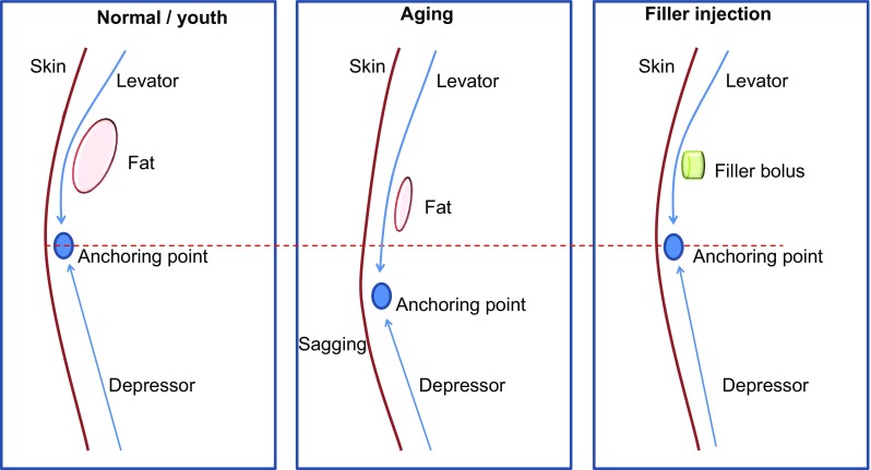
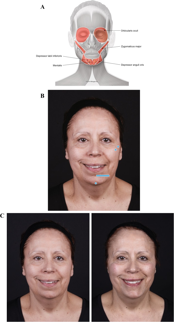
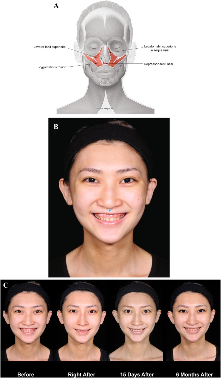
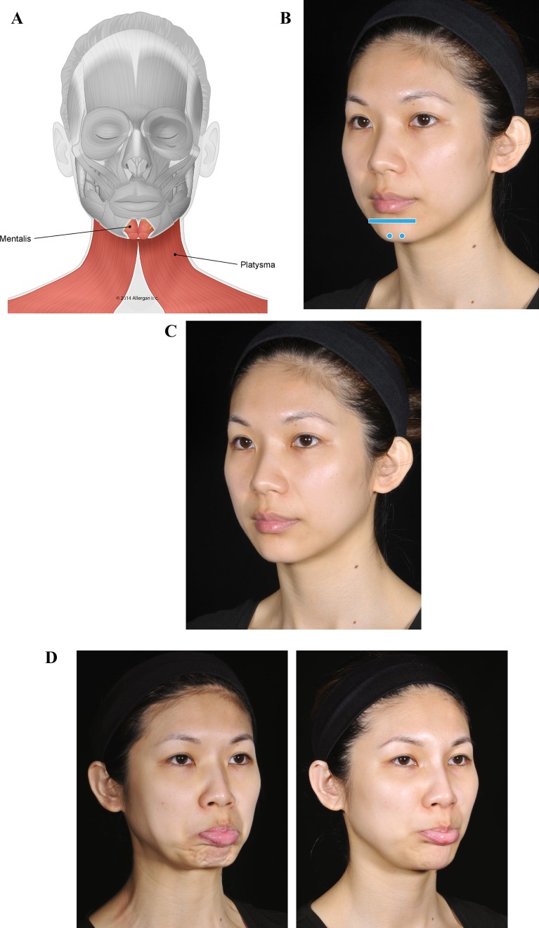
Consideration of facial muscle dynamics is underappreciated among clinicians who provide injectable filler treatment. Injectable fillers are customarily used to fill static wrinkles, folds, and localized areas of volume loss, whereas neuromodulators are used to address excessive muscle movement. However, a more comprehensive understanding of the role of muscle function in facial appearance, taking into account biomechanical concepts such as the balance of activity among synergistic and antagonistic muscle groups, is critical to restoring facial appearance to that of a typical youthful individual with facial esthetic treatments. Failure to fully understand the effects of loss of support (due to aging or congenital structural deficiency) on muscle stability and interaction can result in inadequate or inappropriate treatment, producing an unnatural appearance. This article outlines these concepts to provide an innovative framework for an understanding of the role of muscle movement on facial appearance and presents cases that illustrate how modulation of muscle movement with injectable fillers can address structural deficiencies, rebalance abnormal muscle activity, and restore facial appearance.

Level of evidence v: This journal requires that authors assign a level of evidence to each article. For a full description of these Evidence-Based Medicine ratings, please refer to the Table of Contents or the online Instructions to Authors www.springer.com/00266 .

Keywords: Esthetic facial procedures; Hyaluronic acid; Injectable fillers; Myomodulation; Palsy.


Conflict of interest statement

MDM is an Allergan plc consultant for speaking events and medical education.

Figures

Fig. 1
Fig. 1
Aging and hypothetical treatment effects on muscle action. Normal youthful facial structure (underlying bone and fat) gives muscle fibers convexity, which allows powerful contraction of levator muscles. The levator and depressor muscles are balanced, and the structures are in their normal/youth position. In aging, underlying support for muscle is lost with deflation of facial structure. I believe that the mechanical advantage provided by the lever fulcrum is diminished and the levator loses lifting power to counteract gravity. Muscle fibers are stretched as skin sags. With reduced opposition, the depressor increases in tone over time and pulls facial structures downward in a domino effect. A filler bolus replaces lost structure (fulcrum), increasing mechanical advantage of the levator muscle. The levator’s movement is facilitated. This reduces sagging and balances contraction of the depressor, halting the chain of events triggered by aging
Fig. 2
Fig. 2
Case 1: Asymmetric smile. The patient was treated on both sides at the zygomatic arch and on the chin on her left side only. Voluma was injected at the bone, 0.1 mL at the zygomatic arch on her right side and 0.1 mL at the zygomatic arch and 0.1 mL at the zygomatic eminence on her left, using a 27-g needle to increase the mechanical advantage of the zygomaticus major. Voluma was injected in labiomental angle superficial to the depressor anguli oris (DAO) (0.7 mL) and chin apex (0.3 mL) using a 25-g blunt microcannula and a fanning technique. a Muscles involved. b Injection sites (blue markings; dot = bolus injection, bar = fanning). c Before treatment (left), the patient has a stronger action of zygomaticus major on her right side (zygomatic smile). On her left side, we notice that her zygomatic is weaker and her DAO is stronger compared with her right side, creating a DAO smile. Note that immediately after treatment (right), the corner of her mouth on her left side is lifted and her smile is more symmetrical. The corners of her mouth are more evenly positioned due to facilitation of zygomaticus major muscle bilaterally and reduced contraction of the DAO on the patient’s left side
Fig. 3
Fig. 3
Case 2: Gummy smile. Treatment was a single bolus of Voluma (0.7 mL) injected using a 25-g cannula in the premaxilla area, at the projection of the anterior nasal spine at the level of the bone. a Muscles involved. b Injection sites (blue marking; dot = bolus injection). c Before treatment (left), the lack of support at the premaxilla distorts the nose position and causes a gummy smile. After treatment with Voluma (right; 6 months), the smile is more limited, correcting both the gummy smile and the distortion of the nose. This result was achieved by creating a mechanical obstacle to depressor septi nasi and upper lip levators
Fig. 4
Fig. 4
Case 3: Lack of proper bone support in the chin. The patient was treated with a total of 4 mL of Voluma (1 mL per side into the labiomental angle and 2 mL in the chin apex using a 25-g cannula). a Muscles involved. b Injection sites (blue markings; dot = bolus injection, bar = fanning). c Before treatment, the patient has no apparent structural deficiency at rest. d Pout. Before treatment (left), lack of support in the chin causes mentalis over-contraction and peau d’orange appearance. The platysma is also activated when pouting. Six months after providing a mechanical obstacle to movement with Voluma in her chin (right), the patient is able to protrude her lower lip without skin wrinkling or recruitment of platysma. Blocking over-contraction of the chin also reduces the development of hypertonic platysmal bands
Fig. 5
Fig. 5
Case 4: Voluma was injected into the labiomental angle at the subcutaneous layer, superficial to depressor anguli oris and depressor labii inferioris (1.0 mL per side). Voluma was also injected at the chin apex into the deep fibers of mentalis (0.5 mL per side) using a 25-g cannula in a fanning pattern, and in a bolus pattern using a 27-g needle to reach the supraperiosteal level (0.3 mL per side). a Muscles involved. b Injection sites (blue markings; dot = bolus injection, bar = fanning). c At rest. Before treatment (left), wrinkling of the chin is evident at rest. Improvement in the chin is observed immediately after injection of Voluma in the labiomental angle and chin apex. d Purse (kiss). Before treatment (left), distortion of mentalis muscle during contraction as the patient purses her lips causes extreme wrinkling of the skin on the chin. After providing a mechanical block of mentalis muscle movement with Voluma injected in the labiomental angle and chin apex, the distortion and skin wrinkling are eliminated
Fig. 6
Fig. 6
Case 5: Lack of projection at the chin. Voluma was injected into the labiomental angle, superficial to depressor anguli oris and depressor labii inferioris (0.5 mL per side), and at the chin apex into the deep fibers of mentalis (1 mL) using a 25-g cannula. Juvéderm Ultra Plus was injected in the lip border (1 mL each in cupid’s bow and lip border) using a 27-g needle. a Muscles involved. b Injection sites (blue markings; dot = bolus injection, bar = fanning). c Purse (kiss). Abnormal pursing movement before treatment (left) is due to a lack of support of chin and lips. Six months after treatment in the labiomental angle and chin (right), the patient is able to correctly contract orbicularis oris without deformation. d Pout. Filler injection in the labiomental angle and in the chin also addresses abnormal pouting movement observed before treatment (left). Six months after the injection (right), the patient is able to pout correctly
Fig. 7
Fig. 7
Case 6: Asymmetric smile. Voluma was injected under the zygomaticus major muscle at the level of the bone in his left cheek, 0.1 mL at the zygomatic arch and 0.2 mL at the zygomatic eminence, using a 27-g needle. Voluma was also injected superficial to the depressor anguli oris and depressor labii inferioris in the labiomental angle (1 mL) on his left and deep to the mentalis muscle at the chin apex (0.5 mL) using a 25-g cannula. a Muscles involved. b Injection sites (blue markings; dot = bolus injection, bar = fanning). c At rest. Before treatment (left), there is slight asymmetry at rest with a more pronounced nasolabial fold and protrusion of lower lip. Immediately after treatment (right), the nasolabial fold and the lower lip look more balanced. d Distortion on animation. On the patient’s left side, his nasal flare is in an upward position due to over-contraction of levator labii superioris alaeque nasi (LLSAN) before treatment (left). As a consequence, there is more upper teeth show. Depressor labii inferioris overaction leads to excessive show of lower teeth. Immediately after treatment (right), the smile line is more balanced as both LLSAN and depressor labii inferioris were blocked and zygomaticus major was facilitated. Filler treatment of lip levators and depressors controls distortion both at rest and on animation
Fig. 8
Fig. 8
Case 7: Palsy. Treatment details are described in Table 2. No touch-up treatments were made at return visits. No botulinum toxin was used in this case. a Muscles involved. b Injection sites (yellow markings = injections under the muscle; red markings = injections superficial to the muscle; blue markings = structural injections). c At rest. Before treatment (left), the patient has classical signs of facial palsy on his right side. Deviation of the mouth toward his left side, with a prominent nasolabial fold and narrower left eye, is observed. After treatment, improvement of asymmetry is observed, including better eye symmetry with less scleral show on his right and better alignment of the oral commissures. d Smiling. Before treatment (left), there is a deviation of the oral commissure toward his left hyperkinetic side (normal side). There is also contraction of orbicularis muscle. His right side (facial palsy) presents contraction of the lateral platysmal band, indicating that the facial nerve was not completely damaged. Slight contraction of the zygomaticus major indicates there is residual activity of the zygomatic branch. Immediately after treatment, there is reduction in the upper lateral excursion of the zygomaticus major muscle on his left side. The platysma is contracting on his right side as he tries to smile. There is better positioning of the oral commissure and upper and lower lips on his right side. One to 6 months after treatment, the patient is smiling more symmetrically. His eyes are narrower bilaterally, and platysmal band contraction on his right side is apparent. e Closing the eyes. Before treatment (left), the patient has excessive contraction of the platysma and activation of orbicularis oris. The patient’s right eyebrow is positioned higher relative to the left. Immediately after treatment, the nasolabial fold on his right side is deeper as a result of an increased lever effect on the upper lip levators. Platysma contraction is similar, but more symmetry in the brows is observed. One month after treatment, contraction of zygomatic muscles is facilitated on his right and less recruitment of platysma is observed. Four months after treatment, the position of the eyebrows is improved and orbicularis oculi contraction appears enhanced. The patient is closing his eye with less recruitment of zygomaticus major. At 6 months, no recruitment of lateral muscles is observed. f. Trying to close the eyes without frowning. Before treatment (left), note the scleral show as the patient tries to close his eyes naturally. Abnormal behavior is observed in lateral muscles including orbicularis oris and platysma. Immediately after filler injection, a complete change in muscle behavior is apparent. The patient is able to close his eyes, but there is still some scleral show. No contraction of zygomaticus major muscle is observed. Four months after treatment, the zygomaticus major is even further improved. At 6 months, the patient is able to close his eyes effortlessly, without recruitment of adjacent muscles

References

    1. Kahn DM, Shaw RB. Overview of current thoughts on facial volume and aging. Facial Plast Surg. 2010;26(5):350–355. doi: 10.1055/s-0030-1265024. - DOI - PubMed
    1. Erickson BP, Lee WW, Cohen J, Grunebaum LD. The role of neurotoxins in the periorbital and midfacial areas. Facial Plast Surg Clin North Am. 2015;23(2):243–255. doi: 10.1016/j.fsc.2015.01.010. - DOI - PubMed
    1. Coleman KR, Carruthers J. Combination therapy with BOTOX and fillers: the new rejuvenation paradigm. Dermatol Ther. 2006;19(3):177–188. doi: 10.1111/j.1529-8019.2006.00072.x. - DOI - PubMed
    1. Carruthers J, Carruthers A, Tezel A, Kraemer J, Craik L. Volumizing with a 20-mg/mL smooth, highly cohesive, viscous hyaluronic acid filler and its role in facial rejuvenation therapy. Dermatol Surg. 2010;36(Suppl 3):1886–1892. doi: 10.1111/j.1524-4725.2010.01778.x. - DOI - PubMed
    1. Goldberg RA, Fiaschetti D. Filling the periorbital hollows with hyaluronic acid gel: initial experience with 244 injections. Ophthal Plast Reconstr Surg. 2006;22(5):335–341. doi: 10.1097/01.iop.0000235820.00633.61. - DOI - PubMed