. 2018 Jul;41(7):998-1007.
doi: 10.1007/s00270-018-1936-x. Epub 2018 Mar 16.

Carbon dioxide Angiography-Guided Renal-Related Interventions in Patients with Takayasu Arteritis and Renal Insufficiency

Affiliations

Carbon dioxide Angiography-Guided Renal-Related Interventions in Patients with Takayasu Arteritis and Renal Insufficiency

Sujith Chacko et al. Cardiovasc Intervent Radiol. 2018 Jul.

Abstract

Background: Use of iodinated contrast agents for angiography in patients with renal insufficiency risks further deterioration of renal function and its adverse sequelae.

Objective: To study the effectiveness and safety of carbon dioxide (CO2) angiography in guiding percutaneous renal-related interventions in patients with Takayasu arteritis and renal insufficiency.

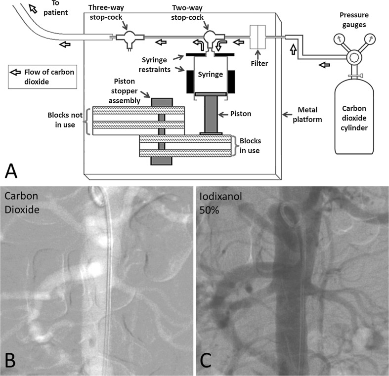
Methods: Data on CO2 angiography-guided interventions were obtained from a 23-year database of 692 Takayasu arteritis patients who underwent percutaneous interventions and were analyzed retrospectively. Follow-up data were also obtained. The CO2 angiography system used was developed in-house and was pressure-driven.

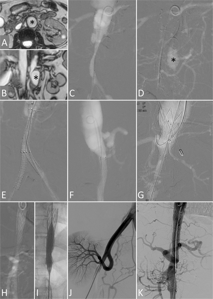
Results: Seven patients (6 female, age 16-59 years, baseline serum creatinine 1.62-4.55 mg/dl, estimated glomerular filtration rate 12.2-36.9 ml/min/1.73 m2) underwent CO2 angiography-guided interventions: five underwent angioplasty or stenting to treat six stenotic/occluded renal arteries, one underwent extensive endovascular repair for spontaneous focal abdominal aortic dissection with false lumen aneurysm and aorto-iliac true lumen narrowing, and one underwent balloon dilatation of previously deployed aortic stents used to treat aortic occlusion at two levels. Follow-up (median 5 years, range 2 months-16 years) was obtained in all patients. All the procedures were successful and resulted in relief of symptoms, better blood pressure control, improvement in left ventricular systolic function and recovery or stabilization of renal function. There were no early or late complications related to CO2 angiography. Three renal lesions that had restenosis at follow-up were managed successfully by repeat intervention.

Conclusion: CO2 angiography-guided renal-related interventions are effective and safe in patients with Takayasu arteritis and renal insufficiency; they significantly improve the care of such patients.

Keywords: Angioplasty; Aortic stenosis; Carbon dioxide; Dissection; Pseudoaneurysm; Renal artery stenosis; Renal failure; Renal insufficiency; Stent; Takayasu arteritis.


Conflict of interest statement

All the authors declare that they have no conflict of interest.

Figures

Fig. 1
Fig. 1
A Diagrammatic representation of the apparatus used for carbon dioxide angiography. Every alternate block of the piston stopper assembly has been left un-shaded to reveal the perpendicular rod around which the blocks can swivel 180°; all the blocks are identical. B, C. Consecutive abdominal aortograms performed in antero-posterior projection in a patient with renal failure using carbon dioxide and 50% iodixanol, respectively, with identical settings of digital subtraction angiography
Fig. 2
Fig. 2
Carbon dioxide-guided renal artery interventions in patients with Takayasu arteritis and renal insufficiency. All images are carbon dioxide angiograms unless stated otherwise. A to C. Case 1. Baseline angiogram (A) shows right renal artery stenosis (arrow). After stent positioning (B) and deployment, the final angiogram (C) showed a good outcome. D to F. Case 2. Baseline angiogram (D) shows ostial left renal artery stenosis (arrow) and long infra-renal aorta narrowing. Renal function normalized after left renal stenting (E). Conventional angiogram (F) obtained 11 years later (5 years after interval infra-renal aortic stenting) shows good long-term outcome. G to I. Case 3. Baseline angiogram (G) shows right renal artery occlusion (black arrow) and left renal artery stenosis (white arrow). Bilateral renal artery stenting was performed (H) leading to normalization of renal function. Conventional angiogram done 4 months later (I) shows good short-term outcome
Fig. 3
Fig. 3
Carbon dioxide-guided aortic and ancillary interventions in patients with Takayasu arteritis and renal insufficiency. A to G. Case 6. Magnetic resonance angiograms in transverse (A) and coronal (B) planes and carbon dioxide angiogram early (C) and late (D) frames in antero-posterior (AP) projection show a single (left) renal artery, focal abdominal aortic dissection, narrowing of the infra-renal aorta and both common iliac arteries and a large false lumen aneurysm on the left lateral aspect of infra-renal aorta (black asterisk). Carbon dioxide AP (E) and lateral (F) angiograms after infra-renal aortic and bilateral iliac artery stenting show relief of stenosis; the superior mesenteric and right renal arteries are not visualized. Carbon dioxide AP angiogram (G) after deployment of a tapered endograft in the abdominal aorta and chimney grafts in the left renal and superior mesenteric arteries shows patency of these arteries; the false lumen aneurysm was no longer visualized in the late frames. H to K. Case 7. Carbon dioxide AP angiogram (H) obtained 7 months after stenting of the lower thoracic-upper abdominal and infra-renal aorta (renal function had deteriorated since then) shows residual stenosis and non-visualization of the renal arteries. The stents were further expanded by balloon dilatation (I), and the right kidney was auto-transplanted (J) resulting is normalization of renal function. Conventional AP aortogram (K) obtained 11 years later shows widely patent aortic stents

References

    1. Hawkins IF, Caridi JG. Carbon dioxide (CO2) digital subtraction angiography: 26 year experience at the University of Florida. Eur Radiol. 1998;8:391–402. doi: 10.1007/s003300050400. - DOI - PubMed
    1. Hawkins IF, Cho KJ, Caridi JG. Carbon dioxide in angiography to reduce the risk of contrast-induced nephropathy. Radiol Clin North Am. 2009;47:813–825. doi: 10.1016/j.rcl.2009.07.002. - DOI - PubMed
    1. Kerr GS, Hallahan CW, Giordano J, Leavitt RY, Fauci AS, Rottem M, Hoffman GS. Takayasu arteritis. Ann Intern Med. 1994;120:919–929. doi: 10.7326/0003-4819-120-11-199406010-00004. - DOI - PubMed
    1. Li J, Li H, Sun F, Chen Z, Yang Y, Zhao J, Li M, Tian X, Zeng X. Clinical characteristics of heart involvement in Chinese patients with Takayasu arteritis. J Rheumatol. 2017 Aug 15. 10.3899/jrheum.161514. Epub ahead of print. - PubMed
    1. Hong S, Ghang B, Kim YG, Lee CK, Yoo B. Long-term outcomes of renal artery involvement in Takayasu arteritis. J Rheumatol. 2017;44:466–472. doi: 10.3899/jrheum.160974. - DOI - PubMed

MeSH terms

LinkOut - more resources