. 2018 Sep;75(18):3411-3422.
doi: 10.1007/s00018-018-2800-5. Epub 2018 Mar 16.

Impact of atypical mitochondrial cyclic-AMP level in nephropathic cystinosis

Affiliations

Impact of atypical mitochondrial cyclic-AMP level in nephropathic cystinosis

Francesco Bellomo et al. Cell Mol Life Sci. 2018 Sep.

Abstract

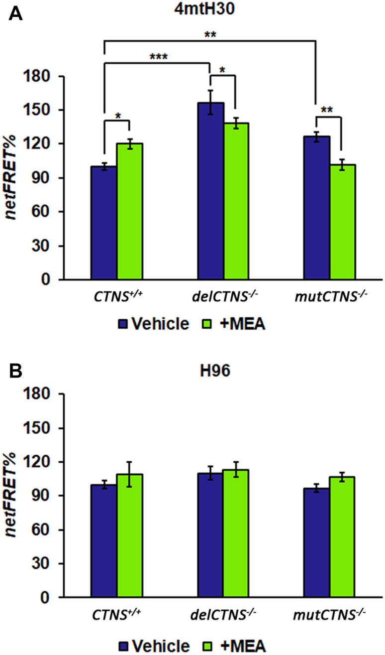
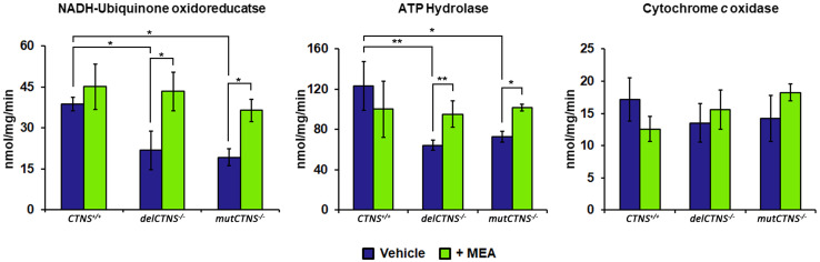
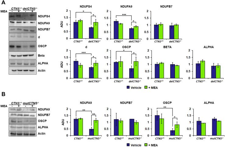
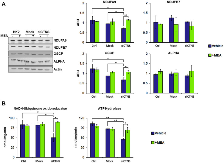
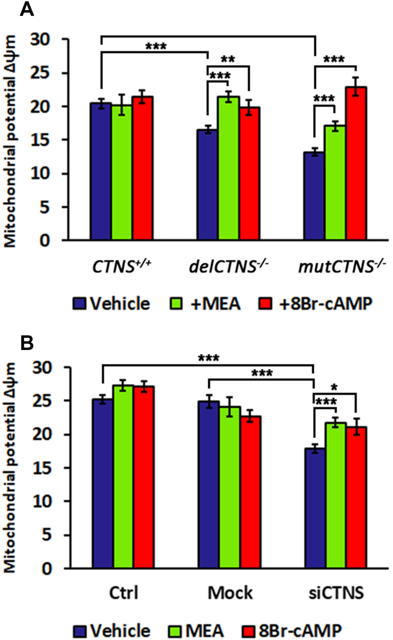
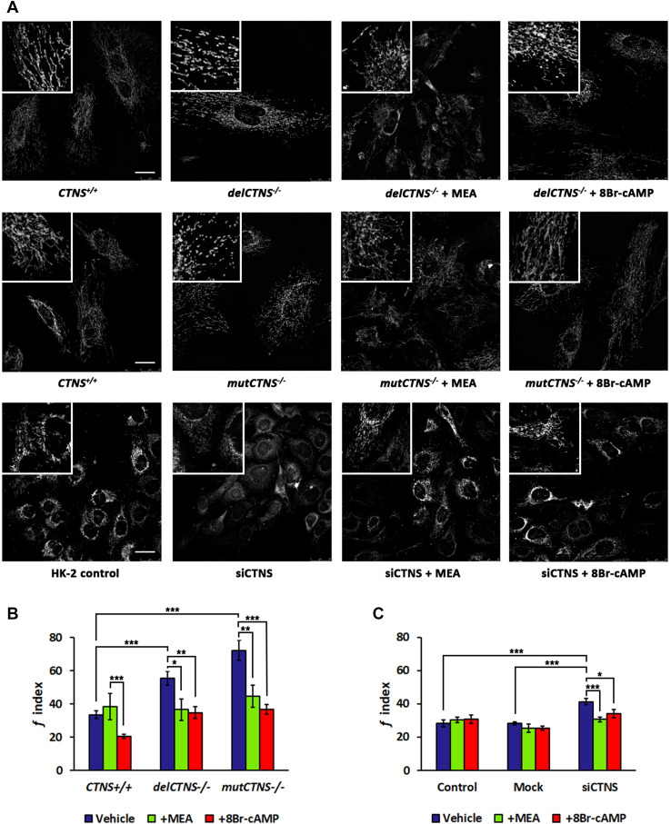
Nephropathic cystinosis (NC) is a rare disease caused by mutations in the CTNS gene encoding for cystinosin, a lysosomal transmembrane cystine/H+ symporter, which promotes the efflux of cystine from lysosomes to cytosol. NC is the most frequent cause of Fanconi syndrome (FS) in young children, the molecular basis of which is not well established. Proximal tubular cells have very high metabolic rate due to the active transport of many solutes. Not surprisingly, mitochondrial disorders are often characterized by FS. A similar mechanism may also apply to NC. Because cAMP has regulatory properties on mitochondrial function, we have analyzed cAMP levels and mitochondrial targets in CTNS-/- conditionally immortalized proximal tubular epithelial cells (ciPTEC) carrying the classical homozygous 57-kb deletion (delCTNS-/-) or with compound heterozygous loss-of-function mutations (mutCTNS-/-). Compared to wild-type cells, cystinotic cells had significantly lower mitochondrial cAMP levels (delCTNS-/- ciPTEC by 56% ± 10.5, P < 0.0001; mutCTNS-/- by 26% ± 4.3, P < 0.001), complex I and V activities, mitochondrial membrane potential, and SIRT3 protein levels, which were associated with increased mitochondrial fragmentation. Reduction of complex I and V activities was associated with lower expression of part of their subunits. Treatment with the non-hydrolysable cAMP analog 8-Br-cAMP restored mitochondrial potential and corrected mitochondria morphology. Treatment with cysteamine, which reduces the intra-lysosomal cystine, was able to restore mitochondrial cAMP levels, as well as most other abnormal mitochondrial findings. These observations were validated in CTNS-silenced HK-2 cells, indicating a pivotal role of mitochondrial cAMP in the proximal tubular dysfunction observed in NC.

Keywords: Cyclic-AMP; Cystinosis; Fanconi syndrome; Lysosomal storage disease; Mitochondria; SIRT3.


Conflict of interest statement

The authors declare no conflicts of interest.

Figures

Fig. 1
Fig. 1
Mitochondrial cAMP level is lower in cystinotic cells and increased by cysteamine treatment. ciPTEC obtained from a healthy volunteer (CTNS+/+) and from two cystinotic patients (delCTNS/ or mutCTNS/) were transfected (transiently) with the EPAC-based FRET sensor target specifically to mitochondria (4mtH30) (a) or cytosol (H96) (b). Where indicated, the cells were treated with 100 µM cysteamine (MEA) or DMSO (vehicle) for 24 h. The histograms represent the means values ± SEM of net% FRET observed (ANOVA test). For other details, see under “Methods” section
Fig. 2
Fig. 2
Activities of complex I and complex V are reduced in cystinotic ciPTEC cells and restored by cysteamine treatment. ciPTEC obtained from a healthy volunteer (CTNS+/+) and from two cystinotic patients (delCTNS/ or mutCTNS/) were treated with 100 µM cysteamine (MEA) or DMSO (vehicle) for 24 h. After incubation, the enzymatic activities of complex I (NADH:ubiquinone oxidoreductase), complex V (ATP hydrolase), and complex IV (cytochrome c oxidase) were performed. The histograms represent the means values ± SD of three independent experiments (Student’s t test). For other details, see under “Methods” section
Fig. 3
Fig. 3
Several subunits of complex I and complex V are reduced in cystinotic ciPTEC cells and restored by cysteamine treatment. a Immunoblotting analysis of ciPTEC obtained from a healthy volunteer (CTNS+/+) and cystinotic patient (delCTNS/). b Immunoblotting analysis of CTNS+/+ and cystinotic patient (mutCTNS/). a, b Where indicated, the cells were treated with 100 µM cysteamine (MEA) or DMSO (vehicle) for 24 h. After incubation, proteins of cellular lysate were loaded on 8% SDS-PAGE, transferred to nitrocellulose membranes, and immunoblotted with the antibodies described in the figure. Protein loading was assessed by reprobing the blots with the actin antibody. Densitometric analysis was performed by VersaDoc imaging system BioRad, using Quantity One software. The histograms represent the means values ± SEM of the ratio of ADU (arbitrary densitometric units) of the immuno-revealed protein normalized on actin level (Student’s t test)
Fig. 4
Fig. 4
HK-2 cells silenced for CTNS gene show a reduction of enzymatic activities and subunit expression levels of the complexes I and V. Cysteamine restores the activities and protein expression. Human kidney-2 cells (HK-2) were transfected with siGENOME human CTNS gene (siCTNS) or non-targeting siRNA (Mock). Where indicated, the cells were treated for 24 h with DMSO (vehicle) or 100 µM cysteamine (MEA). a Proteins of cellular lysate were loaded on 8% SDS-PAGE, transferred to nitrocellulose membranes, and immunoblotted with the antibodies described in the figure. Protein loading was assessed by reprobing the blots with the actin antibody. The histograms represent the means values ± SEM of the ratio of ADU (arbitrary densitometric units) of the immune-revealed protein normalized on actin level (Student’s t test). b Enzymatic activities of complex I (NADH:ubiquinone oxidoreductase) and complex V (ATP hydrolase) were performed as described under “Methods”
Fig. 5
Fig. 5
Mitochondrial membrane potential (Δψm) is lower in cystinotic cells and in HK-2 cells silenced for CTNS gene and rescued by cysteamine or 8Br-cAMP treatment. a Analysis of mitochondrial potential (Δψm) in ciPTEC obtained from a healthy volunteer (CTNS+/+) and from two cystinotic patients (delCTNS/ or mutCTNS/) treated with 100 µM cysteamine (MEA) or 100 µM 8Br-cAMP for 24 h. b Analysis of mitochondrial potential (Δψm) in HK-2 cells transfected with siGENOME human CTNSgene (siCTNS) or non-targeting siRNA (Mock) and treated with 100 µM cysteamine (MEA) or 100 µM 8Br-cAMP for 24 h. Data result from the analysis of the fluorescence intensity mean expressed in arbitrary units (A.U.) ± SEM measured in at least 30 cells (a) and in about 100 cells (b) (Student’s t test)
Fig. 6
Fig. 6
SIRT3 protein level is reduced in cellular models of cystinosis and recovered by cysteamine treatment. Proteins of cellular lysate of the different cellular models were loaded on 8% SDS-PAGE, transferred to nitrocellulose membranes, and immunoblotted with the antibody against SIRT3. Protein loading was assessed by reprobing the blots with the actin antibody. The histograms represent the means values ± SEM of the ratio of ADU (arbitrary densitometric units) of the immune-revealed SIRT3 normalized on actin level (Student’s t test)
Fig. 7
Fig. 7
Cysteamine or 8Br-cAMP treatment ameliorates mitochondrial morphology in cystinotic cells and in HK-2 cells silenced for CTNS gene. a Representative images of mitochondrial network of cells stained with MitoTracker® Orange CMTMRos and a z-stack of optical sections, 10 µm in total thickness, were captured by fluorescence microscopy. Scale bars correspond to 25 µm. b, c Reported the quantification of mitochondrial fragmentation index (f index) in ciPTEC and HK-2 cells treated with 100 µM cysteamine (MEA) or 100 µM 8Br-cAMP for 24 h. Data are the mean values (± SEM) of the analyses performed in cell numbers of n = 20 for ciPTEC cells and n = 30 for HK-2 cells. Significant differences were calculated with Student’s t test

Similar articles

Cited by

References

    1. Acin-Perez R, Salazar E, Kamenetsky M, Buck J, Levin LR, Manfredi G. Cyclic AMP produced inside mitochondria regulates oxidative phosphorylation. Cell Metab. 2009;9:265–276. doi: 10.1016/j.cmet.2009.01.012. - DOI - PMC - PubMed
    1. Ahn BH, Kim HS, Song S, Lee IH, Liu J, Vassilopoulos A, Deng CX, Finkel T. A role for the mitochondrial deacetylase Sirt3 in regulating energy homeostasis. Proc Natl Acad Sci USA. 2008;105:14447–14452. doi: 10.1073/pnas.0803790105. - DOI - PMC - PubMed
    1. Arnold I, Pfeiffer K, Neupert W, Stuart RA, Schagger H. Yeast mitochondrial F1F0-ATP synthase exists as a dimer: identification of three dimer-specific subunits. EMBO J. 1998;17:7170–7178. doi: 10.1093/emboj/17.24.7170. - DOI - PMC - PubMed
    1. Bartsocas CS, Bernstein J, Orloff S, Chandra R, Schulman JD. A familial syndrome of growth retardation, severe Fanconi-type renal disease and glomerular changes–a new entity? Int J Pediatr Nephrol. 1986;7:101–106. - PubMed
    1. Bell EL, Guarente L. The SirT3 divining rod points to oxidative stress. Mol Cell. 2011;42:561–568. doi: 10.1016/j.molcel.2011.05.008. - DOI - PMC - PubMed

MeSH terms

LinkOut - more resources