. 2018 Jun;19(3):223-241.
doi: 10.1007/s10162-018-0662-z. Epub 2018 Mar 16.

Keratinocyte Growth Factor (KGF) Modulates Epidermal Progenitor Cell Kinetics through Activation of p63 in Middle Ear Cholesteatoma

Affiliations

Keratinocyte Growth Factor (KGF) Modulates Epidermal Progenitor Cell Kinetics through Activation of p63 in Middle Ear Cholesteatoma

Tomomi Yamamoto-Fukuda et al. J Assoc Res Otolaryngol. 2018 Jun.

Abstract

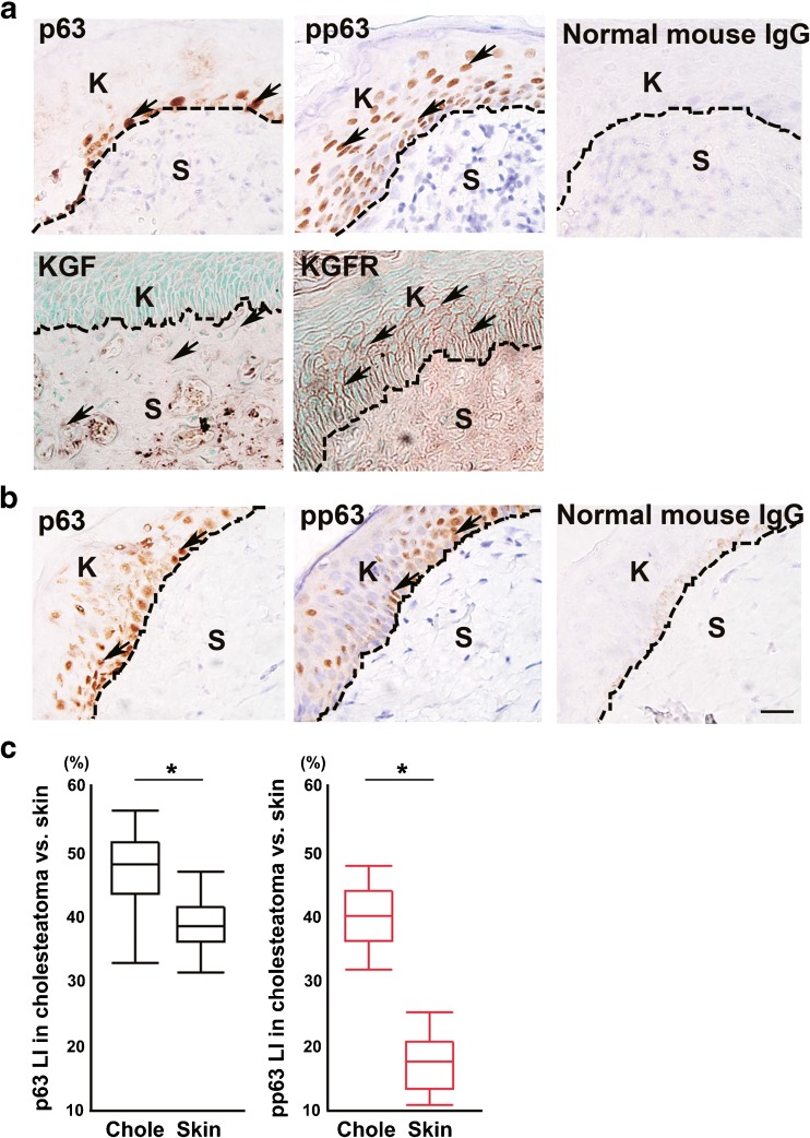
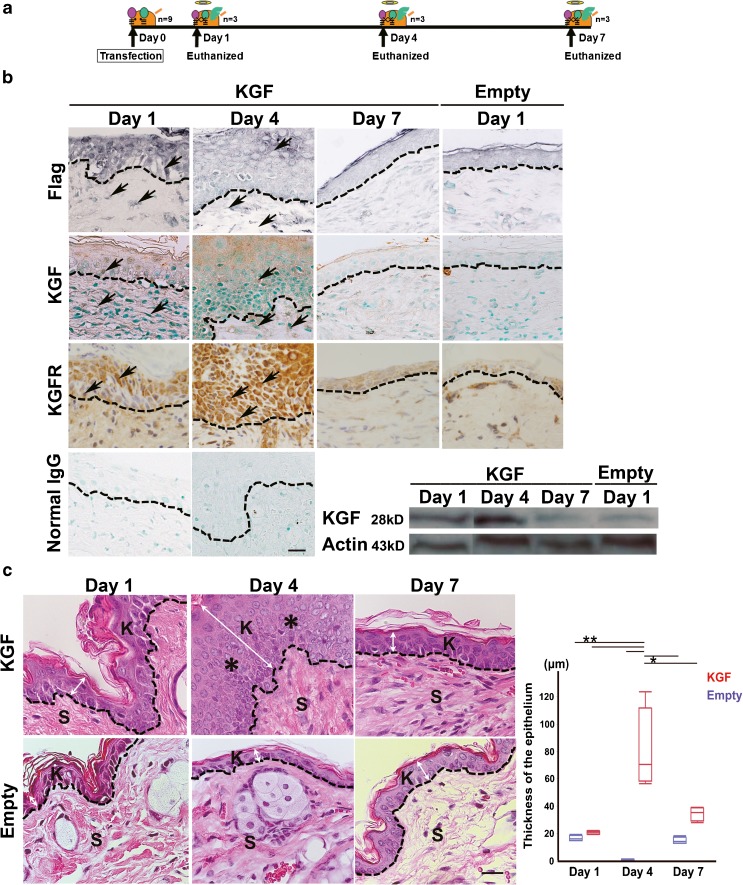
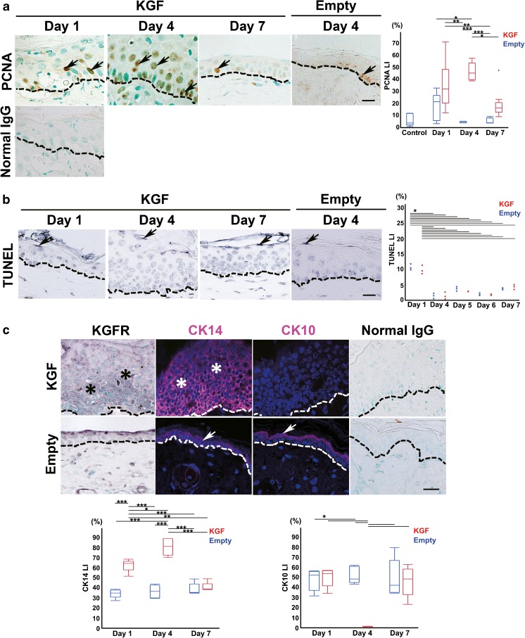
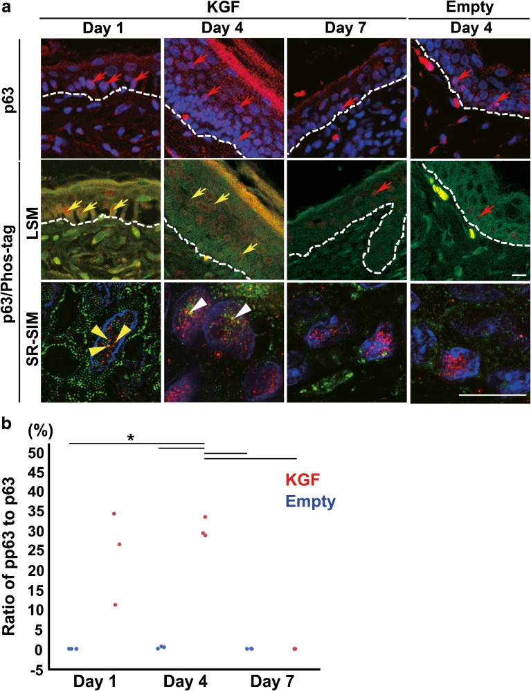
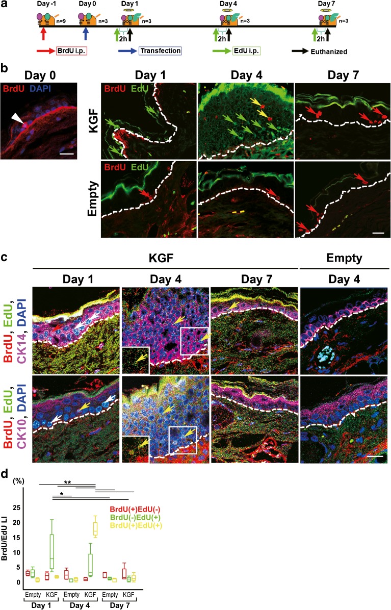
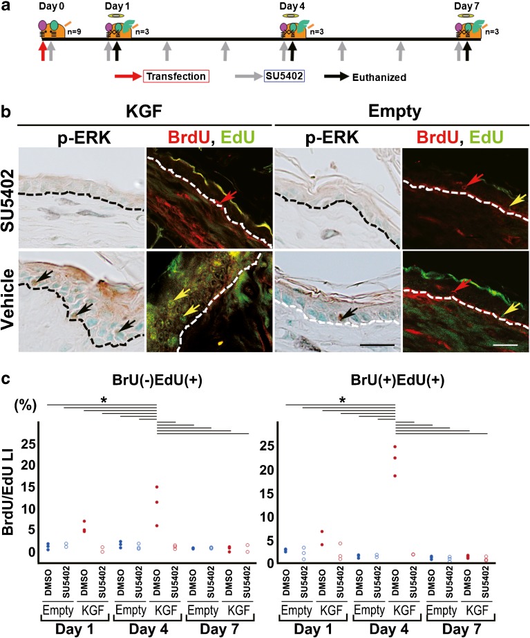
The basal stem/progenitor cell maintains homeostasis of the epidermis. Progressive disturbance of this homeostasis has been implicated as a possible cause in the pathogenesis of epithelial disease, such as middle ear cholesteatoma. In many cases of stem/progenitor cell regulation, the importance of extracellular signals provided by the surrounding cells is well-recognized. Keratinocyte growth factor (KGF) is a mesenchymal-cell-derived paracrine growth factor that specifically participates in skin homeostasis; however, the overexpression of KGF induces middle ear cholesteatoma. In this study, two kinds of thymidine analogs were transferred at different time points and we investigated the effects of overexpressed KGF on the cell kinetics of stem/progenitor cells in vivo. As a result, BrdU(+)EdU(+) cells (stem/progenitor cells) were detected in the thickened epithelium of KGF-transfected specimens. The use of a high-resolution microscope enabled us to analyze the phosphorylated level of p63 in individual nuclei, and the results clearly demonstrated that BrdU(+)EdU(+) cells are regarded as progenitor cells. In the overexpression of KGF, the stimulation of progenitor cell proliferation was inhibited by SU5402, an inhibitor for tyrosine kinase of KGFR. These findings indicate that KGF overexpression may increase stem/progenitor cell proliferation and block terminal differentiation, resulting in epithelial hyperplasia, which is typical in middle ear cholesteatoma.

Keywords: cell tracing; high-resolution microscopy; keratinocyte growth factor (KGF); middle ear cholesteatoma; p63 phosphorylation; progenitor cell.


Conflict of interest statement

Conflict of Interest

This study is supported by a Grant-in-Aid for Scientific Research from the Japanese Society for the Promotor of Science (JSPS) (no. 25462647 and JP16K11186 to T. Yamamoto-Fukuda, no. JP26462608 to N. Akiyama).

Figures

Fig. 1
Fig. 1
Immunohistochemical analysis of KGF, KGFR, p63, and pp63 in human specimens. a p63-positive cells were detected mainly in the basal layer; pp63-positive cells were detected in the basal and upper layers of the cholesteatoma matrix (upper panels). KGF expression was detected in some stromal cells, and KGFR-positive cells were detected in the basal and upper layers of the cholesteatoma matrix (lower panels). Normal mouse IgG was used instead of first antibody as a negative control. b p63-positive cells and pp63-positive cells were detected in the basal and upper layers and the expression level was slightly weak in the control. Normal mouse IgG was used instead of first antibody as a negative control. Arrows indicate positive cells. Dashed lines indicate the basement membrane. K indicates keratinizing squamous epithelium. S indicates the subepithelial region. c Box plot showing the labeling index (LI) of p63 (black boxes) in cholesteatoma (n = 29) vs. skin (n = 22) and pp63 (red boxes) in cholesteatoma (n = 29) vs. skin (n = 21). (p63 LI: *p < 0.0001 as determined by a one-way ANOVA F(1, 49) = 36.22 with a Tukey’s post hoc test, pp63 LI: *p < 0.0001 as determined by a one-way ANOVA F(1, 48) =294.20)
Fig. 2
Fig. 2
Morphological changes in the mouse epidermis after KGF gene transfection. a Schematic description of the method for electroporated transfection of KGF vector in mouse ear skin. The animals were euthanized using an intraperitoneal injection of 200 mg/kg pentobarbital and their ear skin tissue was removed 1, 4, and 7 days after vector transfection. b Immunohistochemical analysis using the anti-FLAG M2 and KGF antibody in sections of KGF gene-transfected ear skin tissue (KGF) (day 1, day 4, and day 7) and the vector alone-transfected ears (Empty) (day 1). Intense staining for FLAG and KGF was detected abundantly in epithelial cells and in some stromal cells of KGF gene-transfected ear skin tissue at day 1 and day 4. FLAG-hKGF expression vector was successfully transfected in vivo and induced KGF expression until day 4. KGFR expression was detected until day 4. No staining of Flag, KGF, and KGFR was observed in the vector alone-transfected ears. No staining was observed with normal mouse IgG and rabbit IgG instead of first antibody. The results of a Western blot analysis of KGF in transfected tissue. KGF: lysate of the skin of the hKGF gene-transfected ears. Empty: lysate of skin of the vector alone-transfected ears. Each lysate was obtained after vector transfection at day 1, day 4, and day 7. The total applied volume was 50 μg per lane. Each lane was reacted with 0.2 μg/ml anti-KGF antibody. An intense band was detected in the hKGF-transfected ears at day 4. c H&E staining of mouse ear skin tissue after vector transfection. KGF gene-transfected ear (KGF) (upper panels, day 1, day 4, and day 7). Vector alone-transfected ear (Empty) (lower panels, day 1, day 4, and day 7). Single transfection of KGF-induced keratin accumulation and thickened squamous epithelium (asterisk) of ear skin tissue at day 4. Dashed lines indicate the basement membrane. K indicates keratinizing squamous epithelium. S indicates the subepithelial region. Scale bar, 20 μm. Arrows indicate positive cells. Double arrow indicates the thickness of the epithelium. Scale bars, 20 μm. Box plot showing the thickness of the epithelium (μm) in vector alone-transfected ear skin (blue boxes) and KGF gene-transfected ears (red boxes). (*p < 0.001, **p < 0.0001 as determined by a two-way ANOVA F(5, 18) = 19.74 with a Tukey’s post hoc test)
Fig. 3
Fig. 3
KGF gene transfection induced epithelial cell proliferation in mouse ear skin tissue. a Immunohistochemical detection of PCNA (a marker of late G1 to S) in sections of KGF gene-transfected ear skin tissue (KGF; day 1, day 4, and day 7) and vector alone-transfected ear skin tissue (Empty; day 4). PCNA-positive cells (arrows) were detected in the basal layer at day 1, basal and upper layers at day 4. No staining was observed with normal mouse IgG instead of first antibody. Dashed lines indicate basement membrane. Scale bar, 20 μm. Box plot showing the PCNA LI of the epithelium in vector alone-transfected ear skin (day 1, n = 7, day 4, n = 4, day 7, n = 4) (blue boxes) and the KGF gene-transfected ears (day 1, n = 6, day 4, n = 7, day 7, n = 10) (red boxes). (*p < 0.001, **p < 0.005, ***p < 0.0001 as determined by a two-way ANOVA F(5, 32) = 11.87 with a Tukey’s post hoc test). b TUNEL-positive epithelial cells in mouse ear skin tissue after vector transfection. TUNEL staining in sections of KGF gene-transfected ear skin tissue (KGF; day 1, day 4, and day 7) and vector alone-transfected ear skin tissue (Empty) (day 4). A few epithelial cells in the cornified layer were TUNEL-positive (arrows). Dashed lines indicate basement membrane. Scale bar, 20 μm. Dots plot showing the TUNEL LI of the epithelium in vector alone-transfected ear skin (n = 3 for each) (blue boxes) and KGF gene-transfected ears (n = 3 for each) (red boxes). (*p < 0.0001 as determined by a two-way ANOVA F(9, 20) = 54.17 with a Tukey’s post hoc test). c Immunohistochemical detection of KGFR, and immunofluorescence detection of CK14 (undifferentiated cell marker) and CK10 (differentiated cell marker) in the section of KGF gene-transfected (KGF, day 4) and vector alone-transfected rat ear skin tissue (Empty, day 4). The expression of KGFR was detected in the basal and upper layers of the KGF gene-transfected epithelium at day 4 but scarcely detected in vector alone-transfected epithelium. CK14 positive cells (magenta) were detected in all layers but CK10-positive cells (magenta) were not detected in any layer at day 4 after KGF gene transfection. In vector alone-transfected epithelium, CK14 was detected in the basal layer and CK10 was detected in the upper layer. As a negative control, sections were reacted with normal rabbit IgG instead of specific antibodies. Arrows: positive cells. Asterisks: positive region. Dashed lines: basement membrane. Scale bars, 20 μm. Nuclei stained with DAPI (4′, 6′- diamidino-2-phenylindole) (blue). Box plot showing the CK14 LI or CK10 LI of the epithelium in vector alone-transfected ear skin (n = 5 for each) (blue boxes) and KGF gene-transfected ears (n = 5 for each) (red boxes). (CK14: *p < 0.05, **p < 0.001, ***p < 0.0001 as determined by a two-way ANOVA F(5, 24) = 46.09 with a Tukey’s post hoc test, CK10: *p < 0.0001 as determined by a two-way ANOVA F(5, 24) = 13.81 with a Tukey’s post hoc test)
Fig. 4
Fig. 4
KGF gene transfection activated the phosphorylation level of p63 in epithelial cells. a Immunofluorescence detection of p63 (red) in the section of KGF gene-transfected (KGF, day 1, day 4, and day 7) and vector alone-transfected mouse ear skin tissue (Empty, day 4) (upper panels). DAPI (4′, 6′- diamidino-2-phenylindole) was used to stain the nuclei (blue). The expression of p63 (red arrows), a reliable stem cell marker of stratified epithelia, was detected in many basal cells and in some upper basal cells at day 1 and in many upper basal cells at day 4 after KGF gene transfection. Double immunofluorescence detection of p63 and Phos-tag in the section of KGF gene-transfected (day 1, day 4, and day 7) (middle panels) and vector alone-transfected (day 4) mouse ear skin tissue (middle panels). Merged LSM images and SR-SIM images show p63 (red) and Phos-tag (green) and nuclei stained with DAPI (blue). In KGF gene-transfected ear skin tissue, high pp63 expression (yellow arrows) within the nuclei was detected in the basal and upper layers at day 1 and in the upper layer at day 4. SR-SIM images (lower panels) show three yellow dots depicting the pp63 (yellow arrow heads) detected in the nuclei of p63-positive basal cells at day 1 and pp63 spots (white arrow heads) in upper-layer cells at day 4 after KGF gene transfection. Dashed lines: basement membrane. Scale bars, 10 μm. b Dots plot showing the ratio of pp63/p63 in a nuclear of the epithelium in vector alone-transfected ear skin (n = 3 for each) (blue dots) and KGF gene-transfected ear (n = 3 for each) (red dots). (*p < 0.0001 as determined by a two-way ANOVA F(5, 12) = 24.96, with a Tukey’s post hoc test)
Fig. 5
Fig. 5
Analyzing the effect of overexpressed KGF on cell kinetics of stem cells, progenitor cells and more differentiated cells using a cell tracing system in vivo. a Schematic description of the method for injecting two thymidine analogues in KGF-transfected mouse ear skin. To investigate the stem cells or progenitor cells after KGF transfection, BdU and ErdU were injected at different time points. BrdU was injected intraperitoneally at 24 h prior to vector transfection and EdU was injected intraperitoneally at 2 h prior to each euthanization. b In the left panel, merged images consisting of BrdU (red) and nuclei stained with DAPI (blue) in the section at day 0. BrdU(+) cells (white arrow head) were detected in the basal layer at day 0. Double immunofluorescence detection of BrdU (red) and EdU (green) was performed in sections of KGF gene-transfected (KGF) and vector alone-transfected ear skin tissue (Empty) (day 1, day 4, and day 7). BrdU(+)EdU(−) cells (red arrow) were detected in the basal layer of KGF gene-transfected ear skin tissue at day 1 and day 7, and vector alone-transfected ear skin tissue. An increased number of BrdU(−) EdU(+) cells (green arrows) were detected in the basal layer at day 1 and upper layer at day 4 after KGF gene transfection. BrdU(+)EdU(+) cells (yellow arrows) were detected in the thickened epithelium in KGF gene-transfected ear skin tissue at day 4. Dashed lines: basement membrane. Scale bars, 20 μm. c Triple immunofluorescence detection of BrdU (red), EdU (green) and CK14 (magenta) or CK10 (magenta) in sections of KGF gene-transfected (KGF) and vector alone-transfected ear skin tissue (Empty) (day 1, day 4 and day 7). Nuclei were stained with DAPI (blue). BrdU(+)EdU(−) cells (red arrow) were detected in the CK14(+)CK10(−) basal layer of KGF gene-transfected ear skin tissue at day 1. An increased number of BrdU(−) EdU(+) cells (white arrows) were detected in the CK14(+)CK10(−) basal layer at day 1 after KGF gene transfection. BrdU(+)EdU(+) cells (yellow arrows) were detected in the CK14(+)CK10(−) thickened epithelium in KGF gene-transfected ear skin tissue at day 4. Inset, high-power view. Dashed lines: basement membrane. Scale bars, 20 μm. d Box plot showing the LI of BrdU(+)EdU(−) positive cells (red boxes), BrdU(−) EdU(+) positive cells (green boxes), or BrdU(+)EdU(+) positive cells (yellow boxes) of the epithelium in vector alone-transfected ear skin (n = 5 for each) and KGF gene-transfected ears (n = 5 for each). BrdU(−) EdU(+) LI (green boxes): *p < 0.01 as determined by a two-way ANOVA F(5, 24) = 5.41 with a Tukey’s post hoc test. BrdU(+)EdU(+) LI (yellow boxes): **p < 0.0001 as determined by a two-way ANOVA F(5, 24) = 139.39 with a Tukey’s post hoc test
Fig. 6
Fig. 6
Under the overexpression of KGF, stimulation of progenitor cell proliferation was inhibited by SU5402, an inhibitor for tyrosine kinase of KGFR. a Schematic description of the method for administering an inhibitor for tyrosine kinase of KGFR (SU5402) in vivo and the injection of two thymidine analogues in KGF-transfected mouse ear skin. BrdU was injected intraperitoneally at 24 h prior to vector transfection and EdU was injected intraperitoneally at 2 h prior to each euthanization. Two millimolars SU5402 was administered via eardrops each day every 24 h from the day of vector transfection to the day before euthanization. b Immunohistochemical detection of p-ERK and double immunofluorescence detection of BrdU (red)/EdU (green) in sections of ear skin tissue treated with SU5402 or 2 % DMSO in PBS (vehicle) after KGF gene transfection (KGF, day 4) or vector-alone transfection (Empty, day 4). The ear tissue treated with SU5402 shows no staining with the anti-p-ERK antibody. In the section of KGF gene-transfected ear tissue treated with a vehicle, many p-ERK-positive cells were stained brown (black arrows). Large numbers of BrdU(+)EdU(+) cells (yellow arrows) were detected in the thickened epithelium of KGF gene-transfected ears treated with a vehicle but not detected in the SU5402-treated specimen. Red arrows: BrdU(+)EdU(−) cells. Scale bars: 20 μm. c Dots plot showing the LI of BrdU(−) EdU(+) positive cells or BrdU(+)EdU(+) positive cells of the ear epithelium treated with SU5402 or 2 % DMSO in PBS (vehicle) after vector-alone transfection (n = 3 for each) or KGF gene transfection (n = 3 for each). BrdU(−) EdU(+) LI: *p < 0.0001 as determined by a two-way ANOVA F(11, 24) = 13.23 with a Tukey’s post hoc test. BrdU(+)EdU(+) LI: *p < 0.0001 as determined by a two-way ANOVA F(11, 24) = 68.55 with a Tukey’s post hoc test

Similar articles

Cited by

References

    1. Akiyama N, Yamamoto-Fukuda T, Takahashi H. Influence of continuous negative pressure in the rat middle ear. Laryngoscope. 2014;124(10):2404–2410. doi: 10.1002/lary.24767. - DOI - PubMed
    1. Andreadis ST, Hamoen KE, Yarmush ML, Morgan JR. Keratinocyte growth factor induces hyperproliferation and delays differentiation in a skin equivalent model system. FASEB J. 2001;15(6):898–906. doi: 10.1096/fj.00-0324com. - DOI - PubMed
    1. Auf demKeller U, Krampert M, Kümin A, Braun S, Werner S. Keratinocyte growth factor: effects on keratinocytes and mechanisms of action. Eur J Cell Biol. 2004;83(11–12):607–612. doi: 10.1078/0171-9335-00389. - DOI - PubMed
    1. Bao S, Wang Y, Sweeney P, Chaudhuri A, Doseff AI, Marsh CB, Knoell DL (2005) Keratinocyte growth factor induces Akt kinase activity and inhibits Fas-mediated apoptosis in A549 lung epithelial cells. Am J Physiol Lung Cell Mol Physiol 288(1):L36–42. doi:0.1152/ajplung.00309.2003 - PubMed
    1. Blanpain C, Simons BD. Unravelling stem cell dynamics by lineage tracing. Nat Rev Mol Cell Biol. 2013;14(8):489–502. doi: 10.1038/nrm3625. - DOI - PubMed

Publication types

MeSH terms

Substances