. 2018 Jun:166:96-108.
doi: 10.1016/j.biomaterials.2018.03.006. Epub 2018 Mar 5.

Production-scale fibronectin nanofibers promote wound closure and tissue repair in a dermal mouse model

Affiliations

Production-scale fibronectin nanofibers promote wound closure and tissue repair in a dermal mouse model

Christophe O Chantre et al. Biomaterials. 2018 Jun.

Abstract

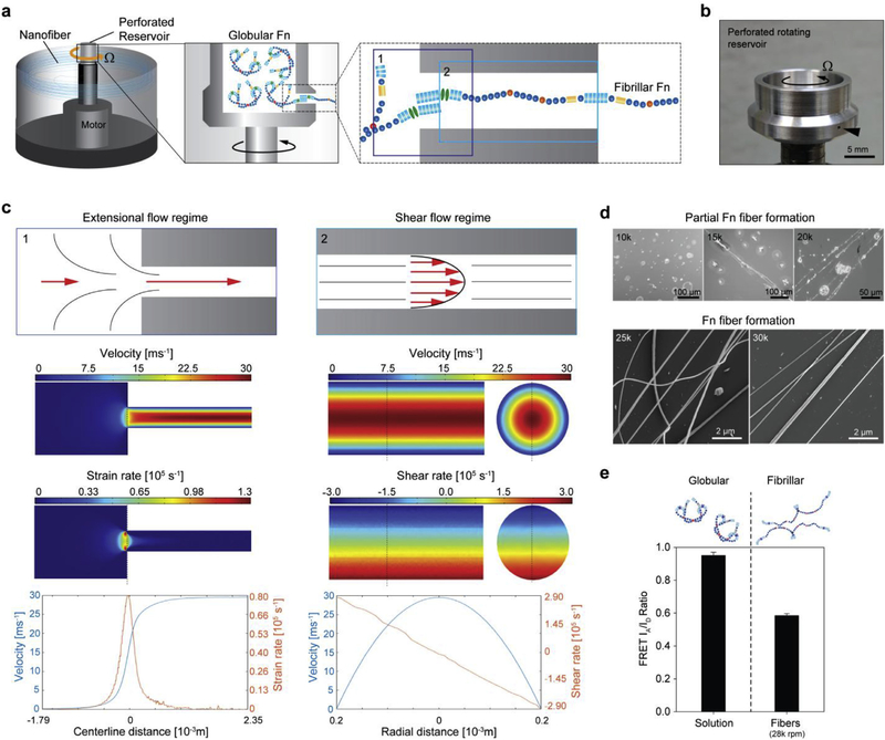
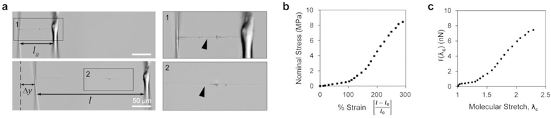
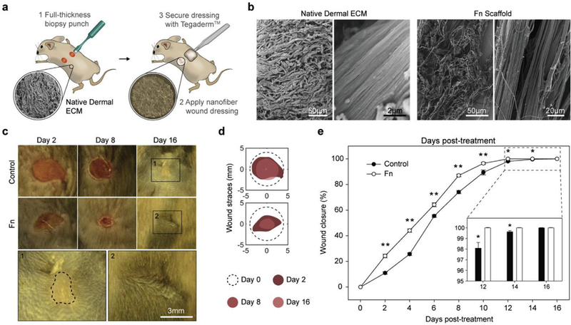
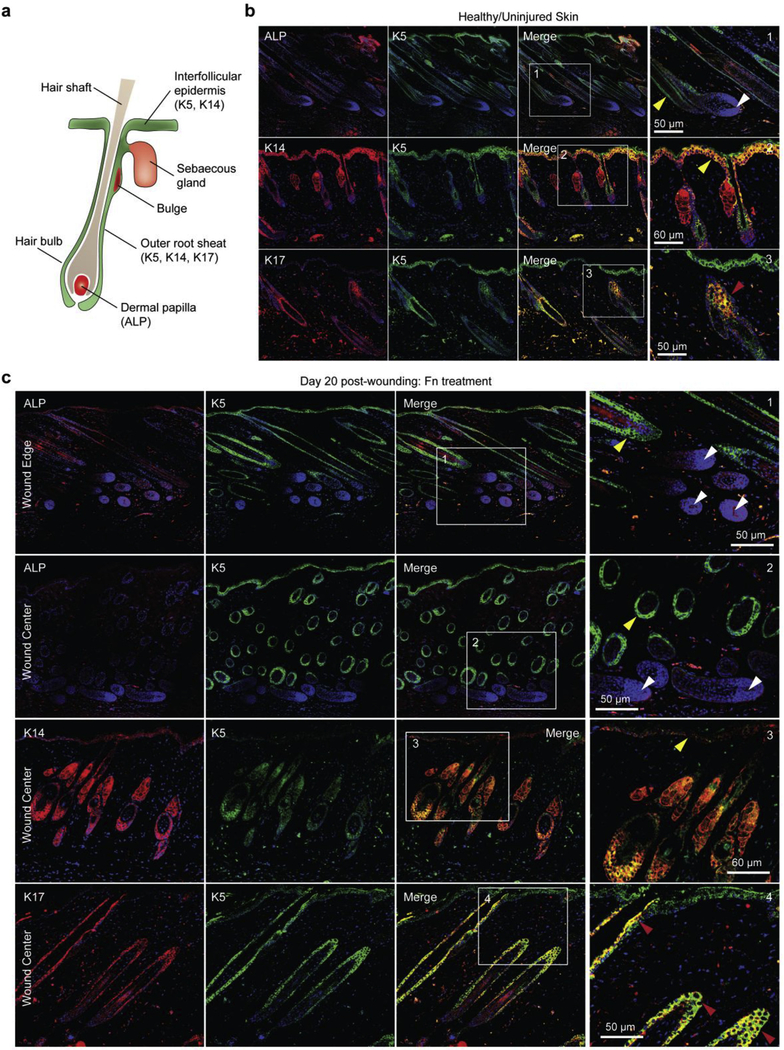
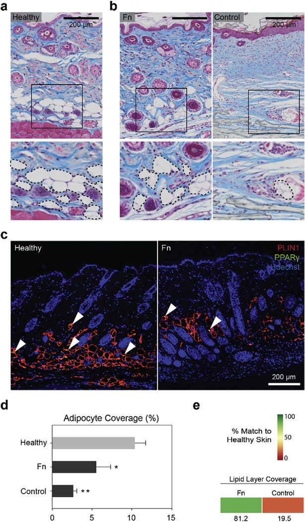
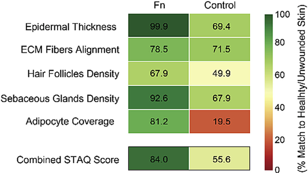
Wounds in the fetus can heal without scarring. Consequently, biomaterials that attempt to recapitulate the biophysical and biochemical properties of fetal skin have emerged as promising pro-regenerative strategies. The extracellular matrix (ECM) protein fibronectin (Fn) in particular is believed to play a crucial role in directing this regenerative phenotype. Accordingly, Fn has been implicated in numerous wound healing studies, yet remains untested in its fibrillar conformation as found in fetal skin. Here, we show that high extensional (∼1.2 ×10⁵ s-1) and shear (∼3 ×10⁵ s-1) strain rates in rotary jet spinning (RJS) can drive high throughput Fn fibrillogenesis (∼10 mL/min), thus producing nanofiber scaffolds that are used to effectively enhance wound healing. When tested on a full-thickness wound mouse model, Fn nanofiber dressings not only accelerated wound closure, but also significantly improved tissue restoration, recovering dermal and epidermal structures as well as skin appendages and adipose tissue. Together, these results suggest that bioprotein nanofiber fabrication via RJS could set a new paradigm for enhancing wound healing and may thus find use in a variety of regenerative medicine applications.

Keywords: Fibrillogenesis; Fibronectin; Hair follicle; Nanofiber; Rotary jet spinning; Wound healing.


Conflict of interest statement

Conflict of Interest

The authors declare no competing financial interests.

Figures

Fig. 1.
Fig. 1.. Hydrodynamic forces produced via rotary jet spinning drive fibrillogenesis of Fn.
(a) The RJS system consists of a perforated reservoir rotating at high speeds. (Insets) Soluble Fn contained in the reservoir is extruded through an orifice and unfolded via centrifugal forces produced by high speed rotation. Insets 1 and 2 show the entry flow and channel flow loci, respectively. (b) Image of the perforated reservoir of the RJS system. (c) Extensional flow regime schematic (left) at the entry shows the Fn solution experiencing high acceleration and high strain rates, depicted with the CFD simulations below. In contrast, the shear flow regime schematic (right) shows the Fn solution experiencing a high velocity and shear gradient across the channel, demonstrated with the CFD simulations below. (d) Scanning electron micrographs (SEM) of Fn spun at different rotation speeds with the RJS. Rotation speeds at 25 k rpm and above show formation of Fn nanofibers, whereas only partial fiber formation is observed at lower speeds. (e) Dual-labeling for FRET shows the reduction in acceptor to donor (IA/ID) ratio before (Fn solution) and after spinning at 28 k rpm. Intensity ratios were 0.95 ± 0.02 and 0.58 ± 0.01 for the Fn solution and the extended fibrillar Fn, respectively. n > 20 measurements per condition.
Fig. 2.
Fig. 2.. Fn nanoflbers extend 300% and exhibit a bimodal stress strain curve.
(a) Differential interference contrast images of a single Fn nanofiber prepared for uniaxial tensile testing (top) and Fn nanofiber during uniaxial tensile testingat ~ 300% strain (bottom). Inset 1 shows a Fn nanofiber (arrowhead) attached to tensile tester μ-pipettes at the resting position, and inset 2 shows Fn nanofiber under uniaxial tension. (b) The stress-strain plot shows that Fn nanofibers produced by RJS have a non-linear behavior, can be characterized by two regimes and can extend up to three times their original length. (c) Results of molecular extension estimation by an eight-chain model.
Fig. 3.
Fig. 3.. Fn nanofiber scaffolds accelerated fiill-thickness wound closure in a C57BL/6 mouse model.
(a) Schematic representation of (1) two full-thickness skin wounds on the back of a mouse performed using a biopsy punch and (2) application of a nanofiber wound dressing. To assure adhesion and stabilization of the nanofibers throughout the study, Tegaderm™ film dressings were applied over the nanofiber scaffolds (3). The control group was likewise covered with a Tegaderm™ film. (b) SEMs of the micro- and macro-structure of native dermal ECM inspired the design and fabrication of Fn scaffolds for optimal integration in the wound. (c) Representative images of the non-treated control group and wounds treated with Fn nanofiber dressings at days 2,8 and 16. Insets bellow show minimal scarring in Fn treatment compared to control (highlighted with the dashed line). (d) From these images, wound edge traces were established for each condition. (e) Normalized wound area over a 16-day period demonstrated that closure rate was significantly increased for Fn dressings compared to the control from day 2 to day 14. Mean and standard error are shown. n = 8 mice and 16 wounds; *p < 0.05 and **p < 0.01 vs. control in a Student’s t-test.
Fig. 4.
Fig. 4.. Fn nanofiber scaffolds promoted native dermal and epidermal architecture recovery.
(a) Masson’s trichrome staining of healthy native tissue sections was performed to establish the design criterion for successful skin tissue restoration. We measured an epidermal thickness of ~20 μm, ECM fiber alignment of ~0.36 (a.u.) as well as ~7 hair follicles and ~3.5 sebaceous glands per surface area of 500 μm2 (c–e). (b) Representative stains of skin tissues with different treatment conditions 20 days post wounding. Black arrowheads indicate original wound edges. Insets demonstrate recovery of epidermal thickness and presence of skin appendages at the center of the wound in the Fn-treated tissue, in contrast with the control group. (c) Epidermal thickness measurements showed that Fn nanofiber dressings restored tissue close to its healthy state, whereas the control had a statistically significant increase in thickness. (d) ECM fiber alignment was used to quantify native tissue (characterized by a basket-woven structure) and scarred tissue (aligned fiber bundles) where 0 is perfectly isotropic and 1 is perfectly anisotropic. Analysis revealed that all recovering tissues were more aligned than native skin, with the Fn condition closer to native skin values than the control. (e) Quantification of hair follicles and sebaceous glands per area demonstrated that Fn wound dressings promoted recovery of skin appendage density close to the native state. This restoration was significantly higher than the control group for both hair follicles and sebaceous glands. Mean and standard error are shown. n = 5–8 wounds; **p < 0.01 vs. Healthy and #p < 0.05, ##p < 0.01 vs. Fn in a one-way ANOVA on ranks with a post hoc multiple comparisons Dunn’s test. (f) To quantify the potency of our treatments, the different parameters measured in c-e were compared to native unwounded tissues and score from 0 to 100% match. Colored boxes are used to represent % match to healthy skin. (g) H&E sections at day 14 post-wounding reveals that wound contraction and hair follicle regeneration are acting synchronously. Wound contraction is observed by a significantly reduced wound size (from 8 mm to −2 mm, black arrowheads in inset), while hair follicle neogenesis is confirmed by the presence of hair follicle pegs growing from the new epidermis at the center of the wound.
Fig. 5.
Fig. 5.. Fn nanofibers restore dermal papillae and recruit basal epithelial cells.
(a) Schematic representation of hair follicle structure with specific markers used in (b–c) labeled. (b) Confocal fluorescent images of alkaline phosphatase (ALP) as well as immunostaining with Keratin 5 (K5), Keratin 14 (K14), Keratin 17 (K17) and DAPI confirmed the presence of dermal papillae (DP) and epithelial cells (EC) in healthy tissues of our mouse model. ECs were observed lining the interfollicular epidermis (IFE) and around the hair follicle shaft (yellow arrowheads). ECs with the K17 marker, specific to the outer root sheath (ORS), were observed in hair follicles only (red arrowheads). White arrowheads highlight the presence of DP (stained with ALP) in the follicle bulb, critical for hair growth and cycling. (c) At day 20 post wounding, K5/K14-positive cells were observed in the IFE and around hair follicles in tissue sections treated with Fn scaffolds. K17-positive cells were observed exclusively in the ORS. ALP-positive cells were observed in re-formed DP, suggestion potential for restoration of functional hair. For the two first panels (ALP/K5 staining), images close to the wound edge (top) and at the center of the wound (bottom) are shown.
Fig. 6.
Fig. 6.. Fn promoted restoration of a lipid layer in the wound.
(a) Masson’s trichrome tissue sections were used to identify presence of the adipose layer in healthy native mouse skin. The inset and dashed lines point out the lipid droplet-carrying adipocytes in the hypodermis of the tissue sections. (b) Representative staining images showing presence of adipocytes in regenerating tissues treated with Fn and the control. (c) PLIN1 and PPARγ immunostaining images of heathy and Fn-treated tissues depicted presence of adipocytes. Arrowheads indicate examples of PPARγ-positive nuclei. (d) Quantitative analysis of Masson’s trichrome images revealed however that both conditions had significantly lower adipocyte area coverage compared to the healthy skin, with a stronger difference calculated in the control condition. n = 5–8 wounds; *p < 0.05, **p < 0.01 vs. Healthy in a one-way ANOVA on ranks with a post hoc multiple comparisons Holm-Sidak test. (e) As previously, treatment conditions were compared to healthy native skin tissue (d) and scored from 0 to 100% match. Colored boxes represent % match to native healthy skin.
Fig. 7.
Fig. 7.. Skin tissue architecture quality (STAQ) index.
The proposed STAQ index quantifies tissue restoration potentiated by the different tested treatments by scoring critical components of skin tissue architecture from 0 to 100% match to healthy/unwounded skin. The Fn score is compared to the no-treatment control. The combined STAQ values indicate an advantage for the Fn dressings with a score above 80% match to native skin. STAQ analysis highlights the improvements still necessary, in particular the ECM Fibers Alignment and Hair Follicles Density parameters with scores below 80% and 70%, respectively.

References

    1. Sen CK, et al., Human skin wounds: a major and snowballing threat to public health and the economy, Wound Repair Regen. 17 (2009) 763–771. - PMC - PubMed
    1. Gurtner GC, Werner S, Barrandon Y, Longaker MT, Wound repair and regeneration, Nature 453 (2008) 314–321. - PubMed
    1. Martin P, Wound healing—aiming for perfect skin regeneration, Science 276 (1997) 75–81. - PubMed
    1. Corr DT, Gallant-Behm CL, Shrive NG, Hart DA, Biomechanical behavior of scar tissue and uninjured skin in a porcine model, Wound Repair Regen. 17 (2009) 250–259. - PubMed
    1. Tomasek JJ, Gabbiani G, Hinz B, Chaponnier C, Brown RA, Myofibroblasts and mechano-regulation of connective tissue remodelling, Nat. Rev. Mol. Cell Biol. 3 (2002) 349–363. - PubMed

Publication types