. 2018 May:518:241-252.
doi: 10.1016/j.virol.2018.03.006. Epub 2018 Mar 15.

Tat controls transcriptional persistence of unintegrated HIV genome in primary human macrophages

Affiliations

Tat controls transcriptional persistence of unintegrated HIV genome in primary human macrophages

Beatrix Meltzer et al. Virology. 2018 May.

Abstract

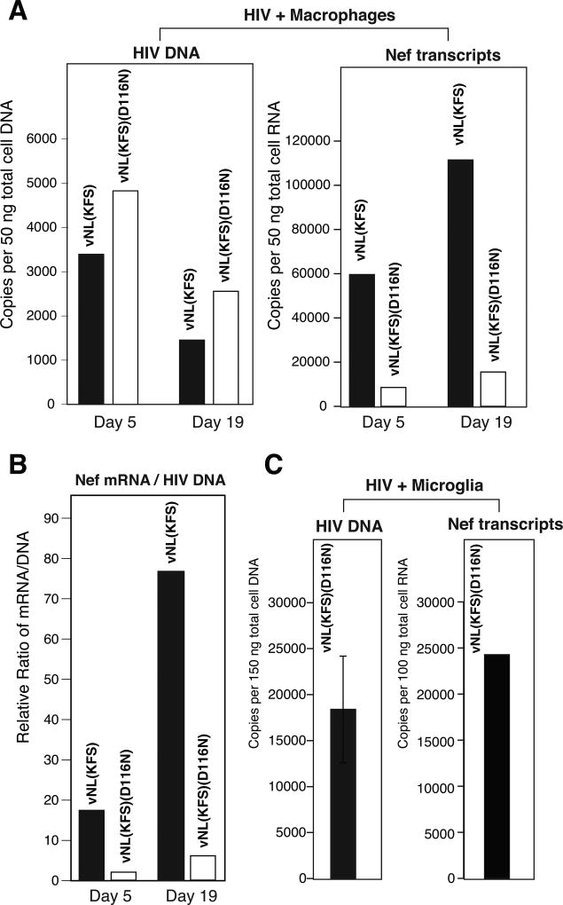
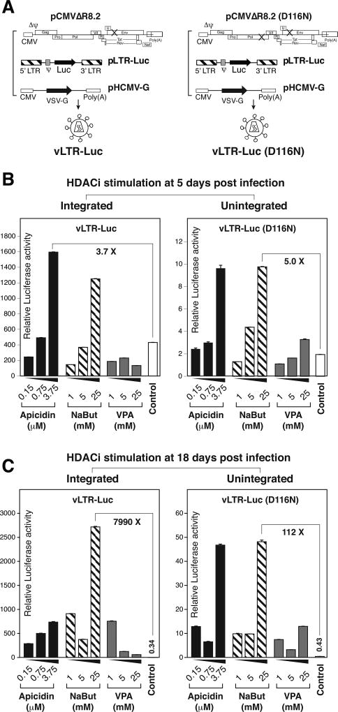
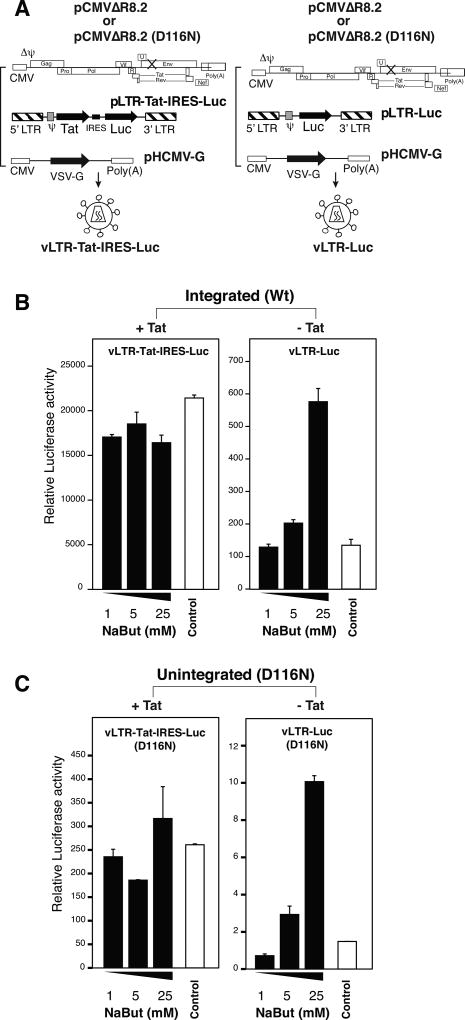
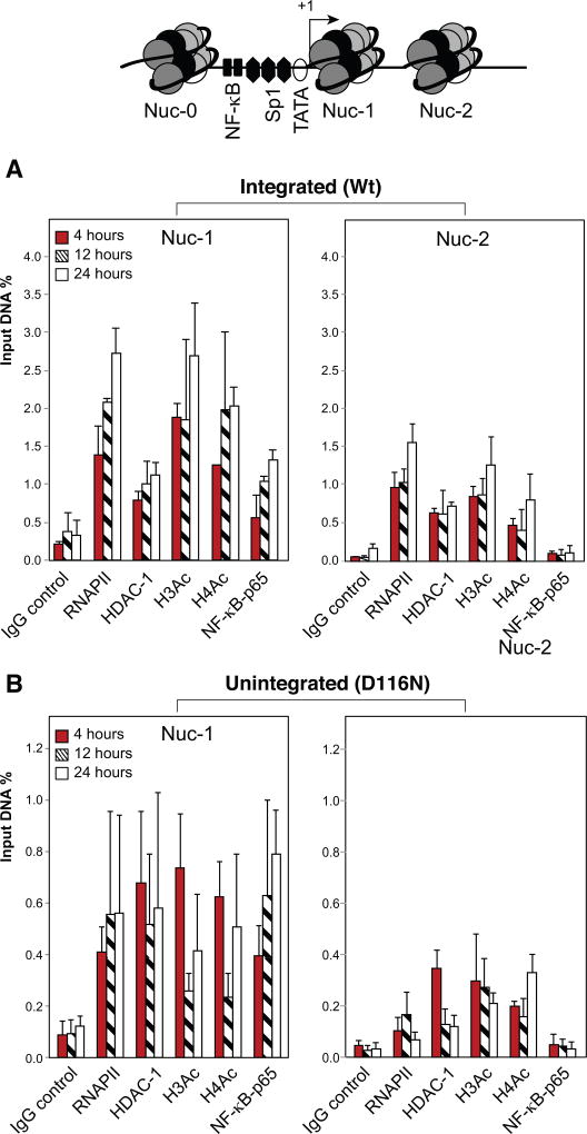
In HIV infected macrophages, a large population of viral genomes persists as the unintegrated form (uDNA) that is transcriptionally active. However, how this transcriptional activity is controlled remains unclear. In this report, we investigated whether Tat, the viral transactivator of transcription, is involved in uDNA transcription. We demonstrate that de novo Tat activity is generated from uDNA, and this uDNA-derived Tat (uTat) transactivates the uDNA LTR. In addition, uTat is required for the transcriptional persistence of uDNA that is assembled into repressive episomal minichromatin. In the absence of uTat, uDNA minichromatin is gradually silenced, but remains highly inducible by HDAC inhibitors (HDACi). Therefore, functionally, uTat antagonizes uDNA minichromatin repression to maintain persistent viral transcription in macrophages. uTat-mediated viral persistence may establish a viral reservoir in macrophages where uDNA were found to persist.

Keywords: HDACi; HIV; Macrophage; Persistence; Reservoir; Tat; Transcription; Unintegrated HIV DNA.


Figures

Fig. 1
Fig. 1. Preintegration transcription occurring at a homogenously low level
(A) An HIV Rev-dependent reporter cell line, Rev-CEM-GFP, was infected with an equal p24 level of HIV-1NL4-3 (Wt) or an integrase mutant, D116N. The percentage of GFP-positive cells was measured at 48 hours post infection with flow cytometry (density plot, left panel). The average mean intensity of GFP in the GFP-positive cells is also shown (histoplot, right panel). (B) The integrase inhibitor, 118-D-24 (50 µM), was used to treat Rev-CEM-GFP cells for 4 hours prior to HIV-1 infection. Cells treated with 118-D-24 or untreated were then infected with an equal p24 level of the Wt virus for 2 hours. Following infection, cells were cultured in the continuous presence of 118-D-24. The percentage (%) and average GFP intensity (M) of GFP-positive cells were measured at 48 hours with flow cytometry. All these experiments have been repeated 3 times.
Fig. 2
Fig. 2. de novo Tat activity from uDNA and its transactivation of preintegration transcription
(A) de novo Tat activity from uDNA. An HIV Tat-dependent indicator cell line, 1G5 cells were infected with the integrate mutant, HIV-1(D116N). For comparison, cells were also pre-treated with the reverse transcriptase inhibitor etravirine. Equal number of cells were lysed and luciferase activity measured at 48 hours post infection. Uninfected 1G5 cells were used as a control. The experiment has been repeated 4 times. (B) Tat-mediated transactivation of preintegration transcription from uDNA. Lentiviral particles with and without the Tat gene (pLTR-GFP-IRES-Tat and pLTR-GFP), and their integrase mutants, pLTR-GFP-IRES-Tat(D116N) and pLTR-GFP)(D116N), were assembled and used to transduce CEM-SS cells. Equal p24 levels of the Tat+ and Tat− viral particles were used. GFP expressions were measured by flow cytometry at 72 hours post infection. The experiments have been repeated 4 times.
Fig. 3
Fig. 3. Different responsiveness of HIV uDNA and provirus to Tat stimulation
(A) Schematic representation of the doxycycline-inducible Tat expression system. HeLa cells (HeLa Tet-On) were stably transfected with a plasmid vector expressing the doxycycline-responsive transcriptional activator, and then transfected with a doxycycline-inducible Tat expression vector, pTRE-Tat86, or a control empty vector, pCDNA3.1. Cells were subsequently infected with a lentiviral particle, vLTR-Luc or its integrase mutant, vLTR-Luc(D116N) (equal p24 for pTRE-Tat86 and pCDNA3.1 transfected cells). Cells were infected with our without Tat-induction by doxycycline. (B) Cells were infected with vLTR-Luc, and luciferase activity was measured at day 1 to 3 post infection. (C) Cells were infected with vLTR-Luc(D116N), and luciferase activity was measured at day 1 to 3 post infection. The mean luciferase activity was from experimental triplicates.
Fig. 4
Fig. 4. Persistent transcription from HIV uDNA in primary macrophages and microglia
(A) Monocyte-derived macrophages were infected with an equal p24 level of HIV-NL(KFS) or its non-integrating mutant HIV-vNL(KFS)(D116N) for 5 and 19 days. Cells were lysed and total cellular DNA and RNA were extracted and used for the quantification of viral DNA and Nef mRNA by real-time PCR and real-time reverse transcriptase PCR. (B) The relative ratio of Nef mRNA and HIV DNA was plotted. (C) Primary human microglia were infected with the non-integrating HIV-NL(KFS)(D116N) for 3 days. Cells were lysed and total cellular DNA and RNA were extracted and used for the quantification of viral DNA and Nef mRNA by real-time PCR and real-time reverse transcriptase PCR.
Fig. 5
Fig. 5. Responses of HIV uDNA to HDAC inhibitor stimulation
(A) Schematics of lentiviral particle assembly. Integrating lentiviral particle vLTR-Luc and its integrase mutant vLTR-Luc(D116N) were assembled by cotransfection with pCMVΔR8.2 or pCMVΔR8.2(D116N), and a VSV-G expression vector, pHCMV-G. Particles were harvested and used to infect monocyte-derived macrophages (MDM) as shown in (B) and (C). (B) At 5 days post infection with vLTR-Luc or vLTR-Luc(D116N), cells were stimulated with apicidin, sodium butyrate (NaBut), and valproic acid (VPA), and harvested for luciferase assay. Control cells were infected but not treated with HDAC inhibitors. HIV viral genomic DNA in cell lysates was quantified for normalization. (C) At 18 days post infection with vLTR-Luc or vLTR-Luc(D116N), cells were identically stimulated and analyzed. The mean luciferase activity was from experimental triplicates.
Fig. 6
Fig. 6. Effects of Tat on HIV uDNA responses to HDACi stimulation
(A) Schematics of lentiviral particle assembly. Integrating lentiviral particles, vLTR-Luc or vLTR-Tat-IRES-Luc, and their integrase mutants, vLTR-Luc(D116N) or Tat-IRES-Luc(D116N), were assembled by cotransfection with pCMVΔR8.2 or pCMVΔR8.2(D116N), and a VSV-G expression vector, pHCMV-G. Particles were harvested and used to infect monocyte-derived macrophages (MDM) as shown in (B) and (C). (B and C) MDMs were infected with an equal p24 level of vLTR-Tat-IRES-Luc or vLTR-Luc, either integrating (B) or non-integrating (C). At 5 days post infection, cells were stimulated with sodium butyrate (NaBut), harvested, and analyzed for luciferase activity. Control cells were infected but not treated with HDAC inhibitors. HIV viral genomic DNA in cell lysates was quantified for normalization. The mean luciferase activity was from experimental triplicates.
Fig. 7
Fig. 7. Assembly of minichromatin onto HIV uDNA in macrophages
U937 cells, a monocytic cell line, were infected with the integration and non-integrating reporter particle vNL-Luc (A) or vNL-Luc(D116N) (B). Cells were cross-linked at 4, 12, and 24 hours post infection, and ChIP assay was performed. On every run, 5% of each sample was analyzed by quantitative real-time PCR to determine the amount of sample immunoprecipitated by individual antibodies. Specific primer sets were used to amplify different regions of the LTR. The reading obtained with preimmune sera was subtracted as background counts.

References

    1. Agbottah E, Deng L, Dannenberg LO, Pumfery A, Kashanchi F. Effect of SWI/SNF chromatin remodeling complex on HIV-1 Tat activated transcription. Retrovirology. 2006;3:48. - PMC - PubMed
    1. Aguilar-Cordova E, Chinen J, Donehower L, Lewis DE, Belmont JW. A sensitive reporter cell line for HIV-1 tat activity, HIV-1 inhibitors, and T cell activation effects. AIDS Res Hum Retroviruses. 1994;10:295–301. - PubMed
    1. Akan E, Chang-Liu CM, Watanabe J, Ishizawa K, Woloschak GE. The effects of vinblastine on the expression of human immunodeficiency virus type 1 long terminal repeat. Leuk Res. 1997;21:459–464. - PubMed
    1. Benkirane M, Chun RF, Xiao H, Ogryzko VV, Howard BH, Nakatani Y, Jeang KT. Activation of integrated provirus requires histone acetyltransferase. p300 and P/CAF are coactivators for HIV-1 Tat. J Biol Chem. 1998;273:24898–24905. - PubMed
    1. Chan CN, Trinite B, Lee CS, Mahajan S, Anand A, Wodarz D, Sabbaj S, Bansal A, Goepfert PA, Levy DN. HIV-1 latency and virus production from unintegrated genomes following direct infection of resting CD4 T cells. Retrovirology. 2016;13:1. - PMC - PubMed

Publication types

Substances

LinkOut - more resources