Review
. 2018 Jun:16:263-275.
doi: 10.1016/j.redox.2018.03.002. Epub 2018 Mar 9.

Diverse roles of mitochondria in ischemic stroke

Affiliations
Review

Diverse roles of mitochondria in ischemic stroke

Jenq-Lin Yang et al. Redox Biol. 2018 Jun.

Abstract

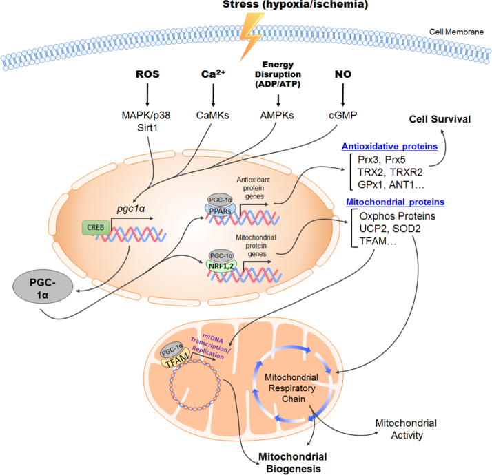
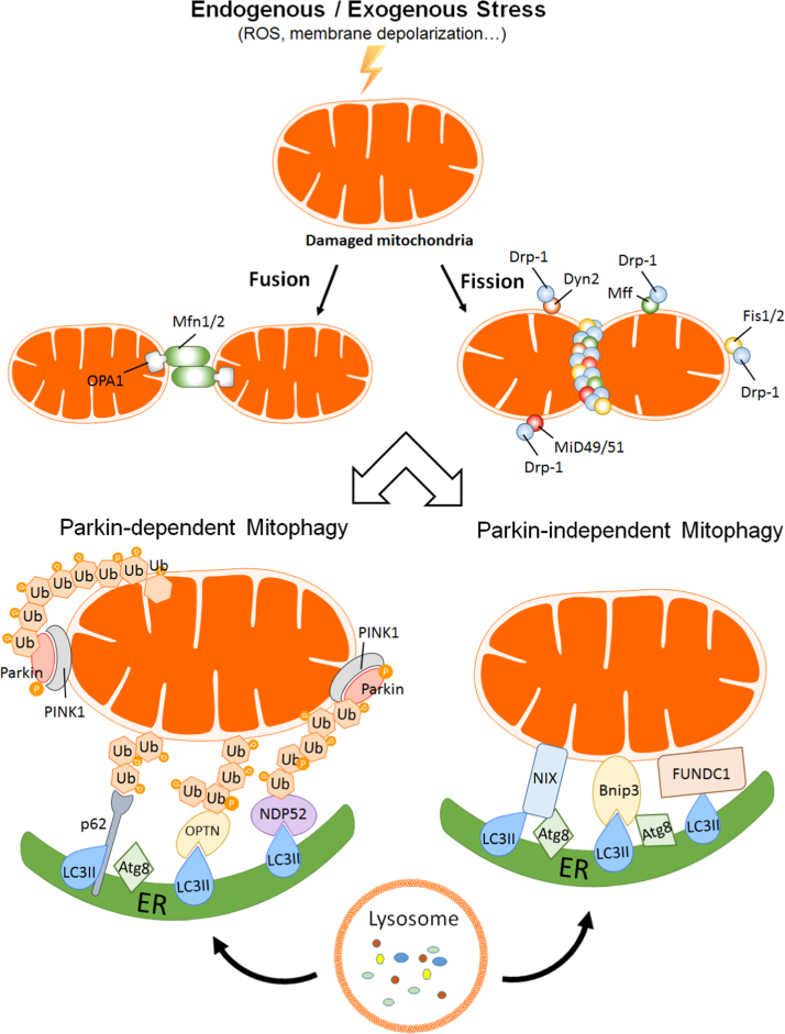
Stroke is the leading cause of adult disability and mortality in most developing and developed countries. The current best practices for patients with acute ischemic stroke include intravenous tissue plasminogen activator and endovascular thrombectomy for large-vessel occlusion to improve clinical outcomes. However, only a limited portion of patients receive thrombolytic therapy or endovascular treatment because the therapeutic time window after ischemic stroke is narrow. To address the current shortage of stroke management approaches, it is critical to identify new potential therapeutic targets. The mitochondrion is an often overlooked target for the clinical treatment of stroke. Early studies of mitochondria focused on their bioenergetic role; however, these organelles are now known to be important in a wide range of cellular functions and signaling events. This review aims to summarize the current knowledge on the mitochondrial molecular mechanisms underlying cerebral ischemia and involved in reactive oxygen species generation and scavenging, electron transport chain dysfunction, apoptosis, mitochondrial dynamics and biogenesis, and inflammation. A better understanding of the roles of mitochondria in ischemia-related neuronal death and protection may provide a rationale for the development of innovative therapeutic regimens for ischemic stroke and other stroke syndromes.

Keywords: Apoptosis; Inflammation; Ischemic stroke; Mitochondria; Mitochondrial biogenesis; Mitochondrial dynamics; Mitophagy.


Figures

fx1
Graphical abstract
Fig. 1
Fig. 1
Pathological signaling pathways involved in mitochondrial function and reactive oxygen species (ROS) generation in the cerebral ischemic cascade. The downstream signaling pathways of ischemic-stroke-induced glutamate excitotoxicity are schematically shown. Excessive Ca2+ influx causes mitochondrial dysfunction and ROS production, leading to various pathological processes, such as mitochondrion-dependent apoptosis, mitochondrial fission and fusion, mitophagy, and DNA damage response and inflammatory responses. Some of these cellular processes eventually lead to cell death. NMDA, N-methyl-D-aspartate; iNOS, inducible NOS; nNOS, neuronal NOS; NO, nitric oxide; NOS, NO synthase.
Fig. 2
Fig. 2
Peroxisome proliferator-activated receptor γ coactivator 1α (PGC-1α) plays a central role in protective mechanisms and mitochondrial biogenesis during hypoxia/ischemia-induced stress. Stress-induced molecules, including reactive oxygen species (ROS), Ca2+, ADP/ATP, and nitric oxide (NO), trigger various signaling pathways and promote PGC-1α expression. Subsequently, PGC-1α, a well-known transcription factor, upregulates the expression of antioxidant proteins and enhances mitochondrial biogenesis to protect neurons against oxidative stress. AMPK, AMP-activated protein kinase; ANT1, adenine nucleotide translocator 1; CAMK, Ca2+/calmodulin-dependent protein kinase; CREB, cAMP response element-binding protein; GPx1, glutathione peroxidase 1; MAPK, mitogen-activated protein kinase; NO, nitric oxide; NRF, nuclear respiratory factor; PPAR, peroxisome proliferator-activated receptor; Prx, peroxiredoxin; SOD2, superoxide dismutase 2; TFAM, transcription factor A, mitochondrial; TRX2, thioredoxin 2; TRXR2, thioredoxin reductase 2; UCP2, uncoupling protein 2.
Fig. 3
Fig. 3
Mitochondrial dynamics and mitophagy have pivotal functions in cell death and survival during cerebral ischemia. Mitochondrial dynamics (fusion/fission) and mitophagy are 2 critical cellular processes maintaining mitochondrial function and energy homeostasis. Mitochondrial fusion and fission maintain functional mitochondria while under a stress insult. However, dysfunctional or overabundant mitochondria continuously undergo mitophagy after mitochondrial fusion of fission. The proper regulation of both mitochondrial dynamics and mitophagy helps cell survival; conversely, imbalanced mitochondrial dynamics and mitophagy or excessive insults lead to cell death. Atg8, autophagy-related protein 8; Bnip3, BCL2/adenovirus E1B 19 kDa protein-interacting protein 3; Dyn, dynamin; Drp1, dynamin-related protein 1; ER, endoplasmic reticulum; Fis1/2, fission protein 1/2; FUNDC1, FUN14-domain-containing protein 1; Mff, mitochondrial fission factor; Mfn, mitofusin; MID49/51, mitochondrial dynamic protein of 49/51 kDa; NDP52, nuclear dot protein of 52 kDa; NIX, Nip3-like protein X; LC3, light chain 3; OPTN, optineurin; PINK1, phosphatase-and-tensin-homolog-induced putative kinase 1; ROS, reactive oxygen species.
Fig. 4
Fig. 4
Mitochondrion-triggered signaling pathways involved in the innate immune response in cerebral ischemic stroke. Mitochondrial reactive oxygen species (mtROS) and DNA (mtDNA) play important roles in triggering the innate immune response via the NLR family pyrin-domain-containing protein 1/3 (NLRP1/3) and p38/NF-κB signaling pathways, respectively. MtROS acts as a critical sensor of inflammation and activates the NLRP1 and NLRP3 inflammasomes, which in turn induces caspase-1 to cleave pro-interleukin (IL)-1β and pro-IL-18, culminating in pyroptotic cell death. Fragmented mtDNA can also be released into the cytosol and activate toll-like receptor 9 (TLR9), triggering the p38 or NF-κB signaling pathways and inducing tumor necrosis factor-α (TNF-α), IL-6, and NLRP1/3 expression. Intravenous immunoglobulin (IVIg) treatment reduces the protein levels of NLRP1 and NLRP3 and inhibits p38 and NF-κB activities, suppressing the downstream inflammatory response. ATF2, activating transcription factor 2; CREB, cAMP response element-binding protein; IKK, IκB kinase; MEF2, myocyte enhancer factor-2; NF-κB, nuclear factor-κB; STAT1, signal transducer and activator of transcription 1.

Similar articles

Cited by

References

    1. Hsieh F.I., Lien L.M., Chen S.T., Bai C.H., Sun M.C., Tseng H.P., Chen Y.W., Chen C.H., Jeng J.S., Tsai S.Y. Get with the guidelines-stroke performance indicators: surveillance of stroke care in the taiwan stroke registry: get with the guidelines-stroke in Taiwan. Circulation. 2010;122:1116–1123. - PubMed
    1. Schwamm L.H., Fonarow G.C., Reeves M.J., Pan W., Frankel M.R., Smith E.E., Ellrodt G., Cannon C.P., Liang L., Peterson E. Get with the guidelines-stroke is associated with sustained improvement in care for patients hospitalized with acute stroke or transient ischemic attack. Circulation. 2009;119:107–115. - PubMed
    1. National Institute of Neurological D., Stroke rt P.A.S.S.G. Tissue plasminogen activator for acute ischemic stroke. N. Engl. J. Med. 1995;333:1581–1587. - PubMed
    1. Jauch E.C., Saver J.L., Adams H.P., Jr, Bruno A., Connors J.J., Demaerschalk B.M., Khatri P., McMullan P.W., Jr, Qureshi A.I., Rosenfield K. Guidelines for the early management of patients with acute ischemic stroke: a guideline for healthcare professionals from the american heart association/american stroke association. Stroke. 2013;44:870–947. - PubMed
    1. Reeves M.J., Arora S., Broderick J.P., Frankel M., Heinrich J.P., Hickenbottom S., Karp H., LaBresh K.A., Malarcher A., Mensah G. Acute stroke care in the us: results from 4 pilot prototypes of the paul coverdell national acute stroke registry. Stroke. 2005;36:1232–1240. - PubMed

Publication types

Substances