. 2018 Jun;1859(6):423-433.
doi: 10.1016/j.bbabio.2018.03.006. Epub 2018 Mar 14.

Estrogen receptor beta modulates permeability transition in brain mitochondria

Affiliations

Estrogen receptor beta modulates permeability transition in brain mitochondria

Suzanne R Burstein et al. Biochim Biophys Acta Bioenerg. 2018 Jun.

Abstract

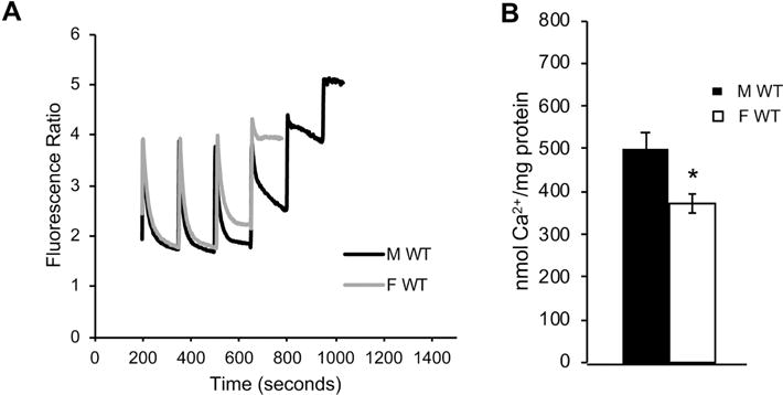
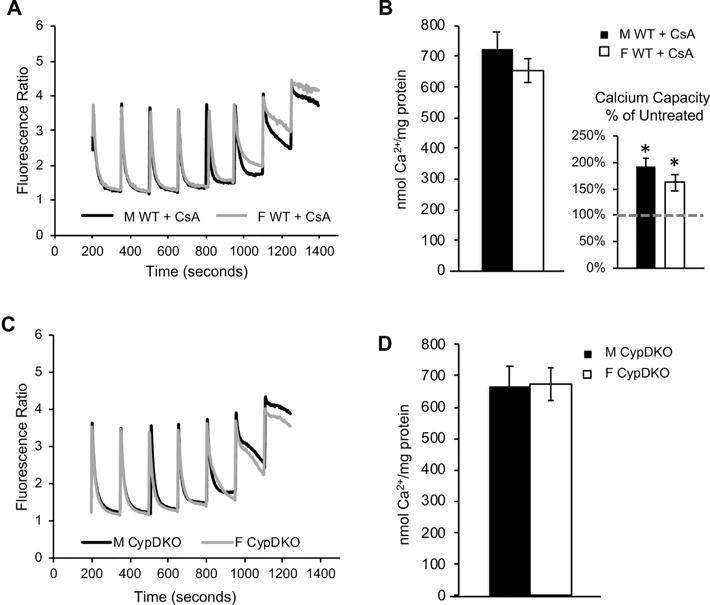
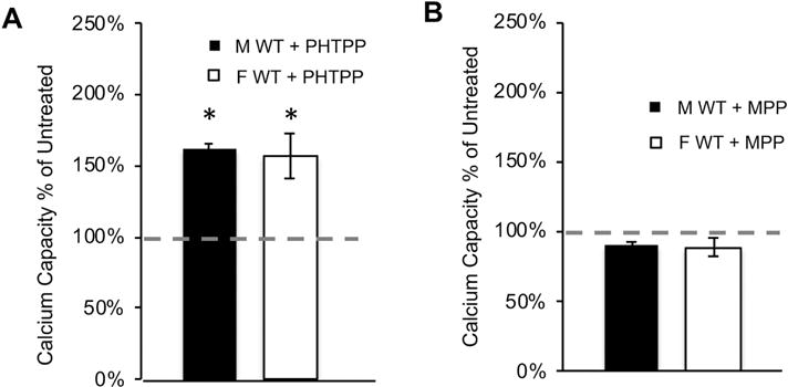
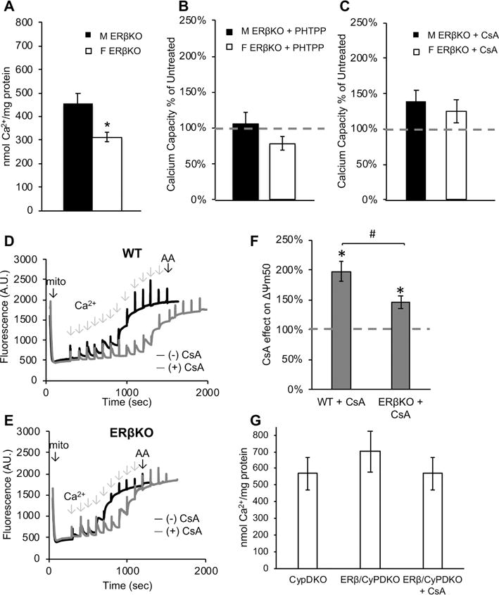
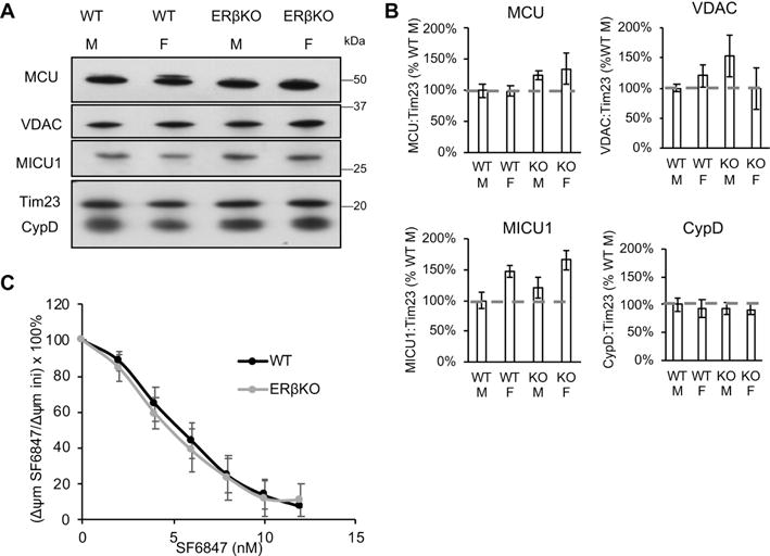
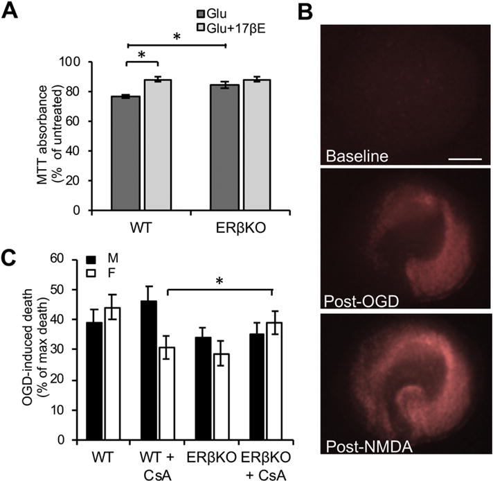
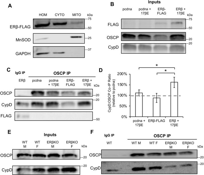
Recent evidence highlights a role for sex and hormonal status in regulating cellular responses to ischemic brain injury and neurodegeneration. A key pathological event in ischemic brain injury is the opening of a mitochondrial permeability transition pore (MPT) induced by excitotoxic calcium levels, which can trigger irreversible damage to mitochondria accompanied by the release of pro-apoptotic factors. However, sex differences in brain MPT modulation have not yet been explored. Here, we show that mitochondria isolated from female mouse forebrain have a lower calcium threshold for MPT than male mitochondria, and that this sex difference depends on the MPT regulator cyclophilin D (CypD). We also demonstrate that an estrogen receptor beta (ERβ) antagonist inhibits MPT and knockout of ERβ decreases the sensitivity of mitochondria to the CypD inhibitor, cyclosporine A. These results suggest a functional relationship between ERβ and CypD in modulating brain MPT. Moreover, co-immunoprecipitation studies identify several ERβ binding partners in mitochondria. Among these, we investigate the mitochondrial ATPase as a putative site of MPT regulation by ERβ. We find that previously described interaction between the oligomycin sensitivity-conferring subunit of ATPase (OSCP) and CypD is decreased by ERβ knockout, suggesting that ERβ modulates MPT by regulating CypD interaction with OSCP. Functionally, in primary neurons and hippocampal slice cultures, modulation of ERβ has protective effects against glutamate toxicity and oxygen glucose deprivation, respectively. Taken together, these results reveal a novel pathway of brain MPT regulation by ERβ that could contribute to sex differences in ischemic brain injury and neurodegeneration.

Keywords: Calcium; Cyclophilin D; Estrogen receptor; Mitochondria; Permeability transition.


Figures

Figure 1
Figure 1. Sex difference in brain mitochondrial calcium capacity
A) Representative fluorimetric traces of calcium uptake in mitochondria purified from male (black line) and female (gray line) adult (130 days of age) mouse forebrain. B) Quantification of brain mitochondrial calcium capacity in wild type males and females. n = 8 animals per group; * p<0.05.
Figure 2
Figure 2. The sex difference in brain mitochondrial calcium capacity is abolished by pharmacological inhibition or genetic ablation of CypD
A) Representative fluorimetric traces of calcium uptake in the presence of CsA (1 μM) in mitochondria purified from male (black line) and female (gray line) adult mouse forebrain. B) Quantification of calcium capacity in male and female brain mitochondria in the presence of CsA. Calcium capacity in CsA treated mitochondria shown as percentage of vehicle treated calcium capacity (dashed line) in the figure inset. * p<0.05 vs. untreated; n = 4 animals per group. C) Representative fluorimetric traces of calcium uptake in mitochondria purified from male (black line) and female (gray line) adult CypDKO mouse forebrain. D) Quantification of brain mitochondrial calcium capacity in CypDKO mice. n = 8 animals per group.
Figure 3
Figure 3. Inhibition of ERβ, but not ERα, increases brain mitochondrial calcium capacity
A) Quantification of calcium capacity in wild type mitochondria treated with the ERβ antagonist PHTPP (5 μM). Data are represented as percent of calcium capacity of respective vehicle-treated mitochondria (dashed line). * p<0.05 vs. untreated; n = 3 animals per group. B) Quantification of mitochondrial calcium capacity of wild type mitochondria treated with ERα antagonist MPP (5 μM). Data are represented as percent of calcium capacity of respective vehicle-treated mitochondria. n = 4 animals per group.
Figure 4
Figure 4. ERβKO brain mitochondria have decreased sensitivity to CsA
A) Quantification of calcium capacity in male and female ERβKO brain mitochondria. * p<0.05; n = 8 animals per group. B) Quantification of mitochondrial calcium capacity of ERβKO mitochondria treated with PHTPP (5 μM). Data are represented as percent of calcium capacity of respective vehicle-treated mitochondria (dashed line). n = 3 animals per group. C) Quantification of mitochondrial calcium capacity of ERβKO mitochondria treated with CsA (1 μM). Data are represented as percent of calcium capacity of respective vehicle-treated mitochondria. n = 6 animals per group. D) Representative trace of Δψm in wild type (WT) brain mitochondria with (gray line) and without (black line) CsA (1 μM). Sequential calcium additions and final addition of antimycin A (AA) are indicated with arrows. E) Representative trace of Δψm in ERβKO brain mitochondria with (gray line) and without (black line) CsA treatment. F) Quantification of half-maximum Δψm in the presence of CsA, expressed as percentage of vehicle-treated, in wild type and ERβKO mitochondria. * p<0.05 vs. vehicle-treated; # p<0.05; n = 6 animals per group. G) Quantification of female brain mitochondrial calcium capacity in CypDKO and ERβ/CypDKO, and ERβ/CypDKO treated with CsA. n = 3 animals per group.
Figure 5
Figure 5. Calcium-related protein expression and bioenergetic capacity of brain mitochondria are not affected by ERβKO
A) Western blot of MCU, VDAC, MICU1, CypD, and Tim23 as mitochondrial protein loading control in wild type (WT) and ERβKO male and female mouse brain mitochondria. B) Quantification of Western blot band immunoreactivity for each protein as a ratio to Tim23 mitochondrial loading control, and expressed as a percentage WT male. n = 3-6 animals per group. C) Depolarization of wild type and ERβKO mitochondria in response to sequential additions (2 nM each) of uncoupler SF6847. Data are calculated as the percentage of the initial membrane potential. n = 4 animals per group.
Figure 6
Figure 6. ERβKO is protective in ischemic injury models
A) Quantification of MTT assay absorbance values in wild type (WT) or ERβKO primary cortical neuronal cultures treated with glutamate (100 μM), with or without 17βE. Data are expressed as percentage of untreated cultures. * p < 0.05 by two-way ANOVA with Bonferroni post-hoc analysis; n = 10-18 independent wells. B) Representative images of hippocampal slice cultures stained with propidium iodide (PI) (indicating cell death) at baseline, 24 hours post-oxygen glucose deprivation (OGD), and 24 hours post-treatment with 1 mM NMDA. C) Quantification of the percentage of PI fluorescence 24 hours post-OGD over the percentage of PI fluorescence post-NMDA treatment, with subtraction of fluorescence prior to any treatment. * p < 0.05 by two-way ANOVA with Bonferroni post-hoc analysis; n = 22-41 slices per group over 8 experiments.
Figure 7
Figure 7. ERβKO decreases the interaction between OSCP and CypD in brain mitochondria
A) Western blot of total homogenate (HOM), cytosolic (CYTO) and mitochondrial (MITO) fractions of cells transfected with ERβ-FLAG. Manganese SOD and GAPDH are used as mitochondrial and cytosolic markers, respectively. B) Western blot of FLAG, OSCP and CypD in enriched mitochondrial fractions from cells expressing empty vector (pcdna) or ERβ-FLAG, with and without 17βE treatment. C) Western blot of co-IP eluate from IgG IP or OSCP co-IP. D) Quantification of the ratio of CypD:OSCP in co-IP eluates. Data are expressed relative to the CypD: OSCP ratio in pcdna transfected cells. * p < 0.05 by one-way ANOVA with Bonferroni post-hoc analysis; n = 7 independent biological replicates. E) Western blot of OSCP and CypD in purified brain mitochondria from wild type (WT) and ERβKO males and females. F) Western blot of co-IP eluate from IgG IP or OSCP co-IP.

References

    1. M. Writing Group. Lloyd-Jones D, Adams RJ, Brown TM, Carnethon M, Dai S, De Simone G, Ferguson TB, Ford E, Furie K, Gillespie C, Go A, Greenlund K, Haase N, Hailpern S, Ho PM, Howard V, Kissela B, Kittner S, Lackland D, Lisabeth L, Marelli A, McDermott MM, Meigs J, Mozaffarian D, Mussolino M, Nichol G, Roger VL, Rosamond W, Sacco R, Sorlie P, Roger VL, Thom T, Wasserthiel-Smoller S, Wong ND, Wylie-Rosett J, C. American Heart Association Statistics, S. Stroke Statistics Heart disease and stroke statistics–2010 update: a report from the American Heart Association. Circulation. 2010;121:e46–e215. - PubMed
    1. Girijala RL, Sohrabji F, Bush RL. Sex differences in stroke: Review of current knowledge and evidence. Vasc Med. 2017;22:135–145. - PubMed
    1. Spychala MS, Honarpisheh P, McCullough LD. Sex differences in neuroinflammation and neuroprotection in ischemic stroke. J Neurosci Res. 2017;95:462–471. - PMC - PubMed
    1. Racay P, Tatarkova Z, Chomova M, Hatok J, Kaplan P, Dobrota D. Mitochondrial calcium transport and mitochondrial dysfunction after global brain ischemia in rat hippocampus. Neurochem Res. 2009;34:1469–1478. - PubMed
    1. Starkov AA, Chinopoulos C, Fiskum G. Mitochondrial calcium and oxidative stress as mediators of ischemic brain injury. Cell Calcium. 2004;36:257–264. - PubMed

Publication types

MeSH terms

Substances