

Response of macrophages in rat skeletal muscle after eccentric exercise
- PMID: 29550244
- PMCID: PMC5911737
- DOI: 10.1016/j.cjtee.2017.12.001
Response of macrophages in rat skeletal muscle after eccentric exercise
Erratum in
-
Erratum regarding missing Declaration of Competing Interest statements in previously published articles.Chin J Traumatol. 2025 Sep;28(5):390. doi: 10.1016/j.cjtee.2020.12.006. Epub 2020 Dec 30. Chin J Traumatol. 2025. PMID: 33386196 Free PMC article. No abstract available.
Abstract
Purpose: Macrophages are known to be important for healing numerous injured tissues depending on their functional phenotypes in response to different stimuli. The objective of this study was to reveal macrophage phenotypic changes involved in exercise-induced skeletal muscle injury and regeneration.
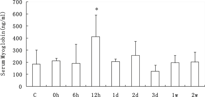
Methods: Adult male Sprague-Dawley rats experienced one session of downhill running (16° decline, 16 m/min) for 90 min. After exercise the blood and soleus muscles were collected at 0 h, 6 h, 12 h, 1 d, 2 d, 3 d, 1 w and 2 w after exercise, separately.
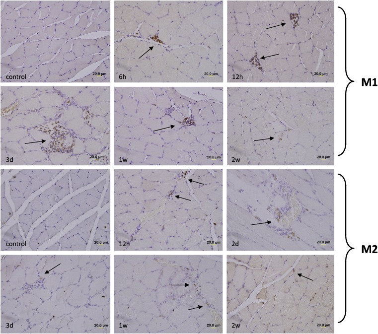
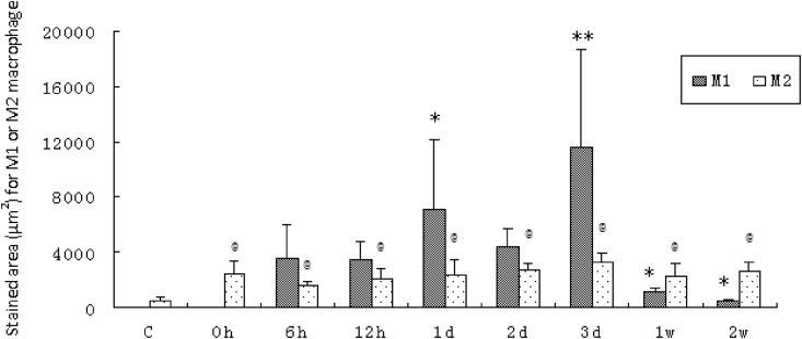
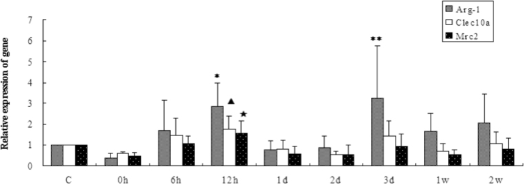
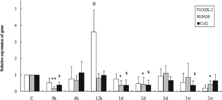
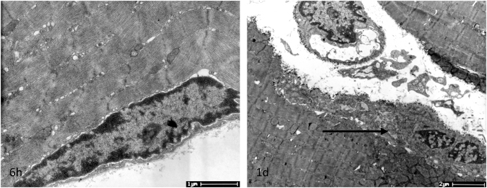
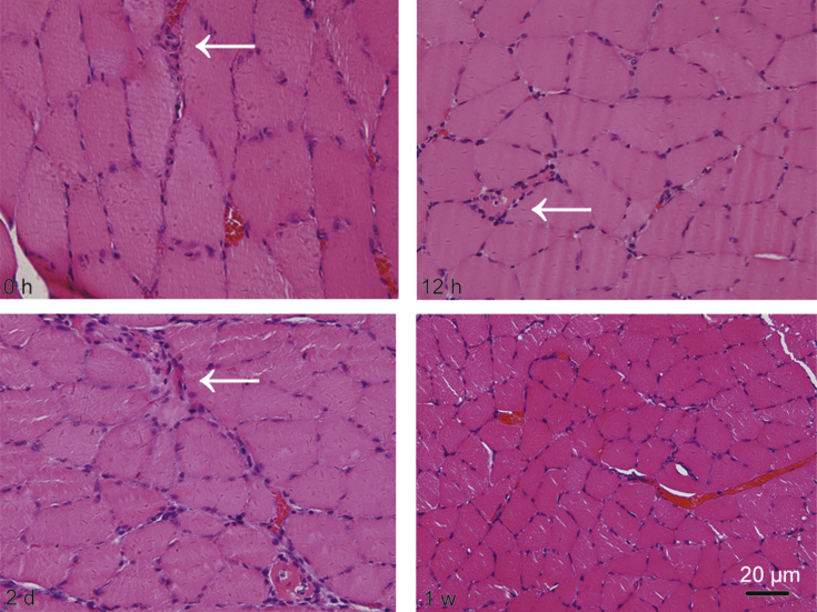
Results: It was showed that CD68+ M1 macrophages mainly infiltrated into muscle necrotic sites at 1-3 d, while CD163+ M2 macrophages were present in muscles from 0 h to 2 weeks after exercise. Using transmission electron microscopy, we observed activated satellite cells 1 d after exercise. Th1-associated transcripts of iNOS and Ccl2 were inhibited post exercise, while COX-2 mRNA was dramatically increased 12 h after running (p < 0.01). M2 phenotype marker Arg-1 increased 12 h and 3 d (p < 0.05, p < 0.01) after exercise, and Clec10a and Mrc2 were up-regulated in muscles 12 h following exercise (p < 0.05, p < 0.05).
Conclusion: The data demonstrate the dynamic patterns of macrophage phenotype in skeletal muscle upon eccentric exercise stimuli, and M1 and M2 phenotypes perform different functions during exercise-induced skeletal muscle injury and recovery.
Keywords: Eccentric exercise; Macrophage phenotype; Muscle injury; Regeneration.
Copyright © 2018 Daping Hospital and the Research Institute of Surgery of the Third Military Medical University. Production and hosting by Elsevier B.V. All rights reserved.
Figures
References
-
- Mantovani A., Biswas S.K., Galdiero M.R. Macrophage plasticity and polarization in tissue repair and remodeling. J Pathol. 2013;229:176–185. - PubMed
-
- Mantovani A., Sica A., Sozzani S. The chemokine system in diverse forms of macrophage activation and polarization. Trends Immunol. 2004;25:677–686. - PubMed
MeSH terms
Substances
LinkOut - more resources
Full Text Sources
Other Literature Sources
Research Materials
Miscellaneous