. 2018 May;265(5):1199-1209.
doi: 10.1007/s00415-018-8830-y. Epub 2018 Mar 17.

Cladribine treatment of multiple sclerosis is associated with depletion of memory B cells

Affiliations

Cladribine treatment of multiple sclerosis is associated with depletion of memory B cells

Bryan Ceronie et al. J Neurol. 2018 May.

Abstract

Background: The mechanism of action of oral cladribine, recently licensed for relapsing multiple sclerosis, is unknown.

Objective: To determine whether cladribine depletes memory B cells consistent with our recent hypothesis that effective, disease-modifying treatments act by physical/functional depletion of memory B cells.

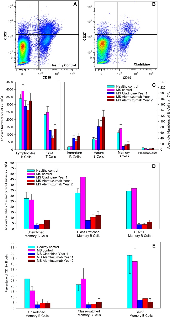
Methods: A cross-sectional study examined 40 people with multiple sclerosis at the end of the first cycle of alemtuzumab or injectable cladribine. The relative proportions and absolute numbers of peripheral blood B lymphocyte subsets were measured using flow cytometry. Cell-subtype expression of genes involved in cladribine metabolism was examined from data in public repositories.

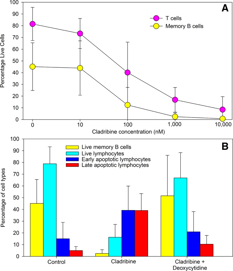
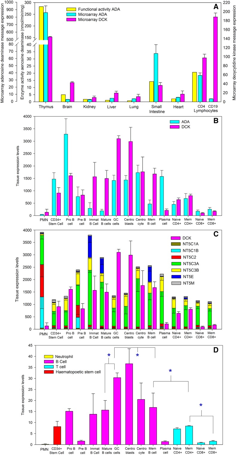
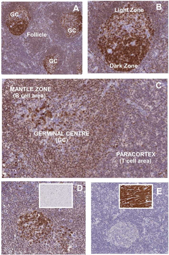
Results: Cladribine markedly depleted class-switched and unswitched memory B cells to levels comparable with alemtuzumab, but without the associated initial lymphopenia. CD3+ T cell depletion was modest. The mRNA expression of metabolism genes varied between lymphocyte subsets. A high ratio of deoxycytidine kinase to group I cytosolic 5' nucleotidase expression was present in B cells and was particularly high in mature, memory and notably germinal centre B cells, but not plasma cells.

Conclusions: Selective B cell cytotoxicity coupled with slow repopulation kinetics results in long-term, memory B cell depletion by cladribine. These may offer a new target, possibly with potential biomarker activity, for future drug development.

Keywords: B cell; Cladribine; Deoxycytidine kinase; Disease-modifying treatment; Memory B cells; Multiple sclerosis.


Conflict of interest statement

Conflicts of interest

GG has received fees for participation in advisory board and speaker fees for Merck. KS has received honoraria and meeting support from Merck. However, Merck was not involved in this study design or its implementation. Other disclosures are considered not relevant but: BC has nothing to declare, BMJ has nothing to declare; DB is a shareholder and consultant to Canbex therapeutics and has received research support from Sanofi-Genzyme; ND has nothing to declare; ZM has nothing to declare; GG has received fees for participation in advisory board from AbbVie Biotherapeutics, Biogen, Canbex, Ironwood, Novartis, Merck, Roche, Sanofi-Genzyme, Synthon, Teva and Vertex; speaker fees from AbbVie, Biogen, Bayer HealthCare, Genzyme, Sanofi-Aventis and Teva. Research support from Biogen, Genzyme, Ironwood, Merck, Merck Serono, Novartis and Takeda. KS has been a PI of trials sponsored by Novartis, Roche and Teva and involved in trials sponsored by Biogen, Sanofi-Genzyme, BIAL, Cytokinetics, and Canbex and has received honoraria and meeting support from Biogen, Novartis, and Teva.

Ethical standard statement

The study was approved by the Health and Social Care Research Ethics Committee B and the Health Research Authority, UK. People were recruited following informed consent. Purchased blood samples were collected with informed consent and did not require additional ethical review.

Figures

Fig. 1
Fig. 1
Cladribine induced lymphocyte killing in vitro. Peripheral blood mononuclear cells were incubated with various concentrations of cladribine for 70 h and were stained with Annexin V and DAPI to detect apoptotic and live cells and were phenotyped with CD3, CD19 and CD27 immunofluorescence using flow cytometry. a Cell viability and different cell subtypes and b Inhibition of 1 μM cladribine-induced lymphocyte cytotoxicity (Live = annexin V−, DAPI−; early = Annexin V+ , DAPI−; Late apoptosis = Annexin V+, DAPI+) in the presence or absence of 250 μM deoxycytidine. Results represent the mean ± standard deviation, n = 6
Fig. 2
Fig. 2
Cladribine induces marked memory B cells depletion. People with MS were treated with either subcutaneous cladribine of alemtuzumab and blood samples were taken at the last blood-screen prior to retreatment at year 1 (cladribine and alemtuzumab year 1) and 12 months after the second cycle (alemtuzumab year 2). Cells were stained with T and B cell markers assessed by flow cytometry and the results represent the mean ± standard deviation (n = 8/group). Flow cytometry of CD19 and CD27 staining in a a healthy control and b a person with MS treated with cladribine. c Total numbers of (left panel) T cells and (right panel) B cell subsets, d total numbers and e percentage of CD19+ B cells of immunoglobulin class-switched (IgD) and unswitched (IgD+) and interleukin 2 receptor expressing memory B cells CD19+, CD27+ memory B cells
Fig. 3
Fig. 3
Microarray expression of purine salvage pathway genes indicates a B cell sensitivity to cladribine. Publically available microarray expression data (http://www.biogps.org) was extracted from the a Geneatlas U133, gcrma and bd Primary cell Atlas. DBS_00013. a Microarray detected gene expression of adenosine deaminase (ADA. 204639_at) and deoxycytidine kinase (DCK. 203303_at) in various tissues in the Geneatlas U133, gcrma. Identifier GSE1133 (http://www.biogps.org). The results represent the mean ± SD in duplicate samples. This was compared to the distribution of function protein expression reported previously [14]. bd The data represent the mean ± SD expression Z scores from: neutrophils (n = 4), CD34+ hematopoietic stem cells (n = 6), Pro-B (n = 2), Pre B (n = 2), immature B cells (Immat, n = 3) and tonsillar mature cells (n = 3), germinal centre cells (GC cells, n = 4), centroblasts (n = 4), centrocytes (n = 4), memory B cells (mem, n = 3) and plasma cells (n = 3), naïve and effector memory (Mem, CCR7, CD45RO+) CD4+ (n = 5/group) and CD8+ T cells (n = 4/group). The expression of a ADA (204639_at) and DCK (23302_at). b The expression of DCK and 5′NT detecting by: NT5C1A (224549_s_at), NT5C1B (243100_at), NT5C2 (209155_s), NT5C3A (225044_at), NT5C3B (209155_s_at), NT5E (203939_at) and NT5M (219708_at). c Expression ratio of DCK expression divided by expression score of NT5C1A and NT5C1B 5′NT that can dephosphorylate adenosine/monophosphate. *Significantly different between groups (P < 0.05)
Fig. 4
Fig. 4
Deoxycytidine kinase expression in lymph node tissue. DCK expression was immunostained in three (ad) people showing: a DCK expression in follicle and secondary follicles containing high-intensity staining in the germinal centres (GC). b Intense immunostaining was present notably in the germinal centre B cells and centroblasts in the dark zone, compared to the light zone contains centrocytes, memory B cells and plasma cells. c Enhanced staining in secondary follicles compared to the paracortex. d DCK expression in lymph nodes with heart myocyte staining in the inset. e NT5C1A expression in lymph nodes with heart myocyte staining in the inset. Elevated message was detected in skeletal and heart muscle compared to peripheral blood mononuclear cells in GEO identifier GDS 3113. Immunostaining is reproduced under the Creative Commons Attribution-Share Alike 4.0 international licence. Images, including description of tissue donor, available from http://V17.proteinatlas.org, https://www.proteinatlas.org/ENSG00000116981-NT5C1A/tissue/lymph+node#img, https://www.proteinatlas.org/ENSG00000116981-NT5C1A/tissue/heart+muscle#img, https://www.proteinatlas.org/ENSG00000156136-DCK/tissue/lymph+node#img and https://www.proteinatlas.org/ENSG00000156136-DCK/tissue/heart+muscle

References

    1. Legroux L, Arbour N. Multiple sclerosis and T lymphocytes: an entangled story. J Neuroimmune Pharmacol. 2015;10:528–546. doi: 10.1007/s11481-015-9614-0. - DOI - PMC - PubMed
    1. Sefia E, Pryce G, Meier UC, et al. Depletion of CD20 B cells fails to inhibit relapsing mouse experimental autoimmune encephalomyelitis. Mult Scler Relat Disord. 2017;14:46–50. doi: 10.1016/j.msard.2017.03.013. - DOI - PubMed
    1. van Oosten BW, Lai M, Hodgkinson S, et al. Treatment of multiple sclerosis with the monoclonal anti-CD4 antibody cM-T412: results of a randomized, double-blind, placebo-controlled, MR-monitored phase II trial. Neurology. 1997;49:351–357. doi: 10.1212/WNL.49.2.351. - DOI - PubMed
    1. Baker D, Marta M, Pryce G, et al. Memory B cells are major targets for effective immunotherapy in relapsing multiple sclerosis. EBioMedicine. 2017;16:41–50. doi: 10.1016/j.ebiom.2017.01.042. - DOI - PMC - PubMed
    1. Cohen JA, Coles AJ, Arnold DL, et al. Alemtuzumab versus interferon beta 1a as first-line treatment for patients with relapsing-remitting multiple sclerosis: a randomised controlled phase 3 trial. Lancet. 2012;380:1819–1828. doi: 10.1016/S0140-6736(12)61769-3. - DOI - PubMed

MeSH terms