. 2018 Jan 1:17:1533033818762910.
doi: 10.1177/1533033818762910.

Construction, Expression, and Characterization of rSEA-EGF and In Vitro Evaluation of its Antitumor Activity Against Nasopharyngeal Cancer

Affiliations

Construction, Expression, and Characterization of rSEA-EGF and In Vitro Evaluation of its Antitumor Activity Against Nasopharyngeal Cancer

Xueting Liu et al. Technol Cancer Res Treat. .

Abstract

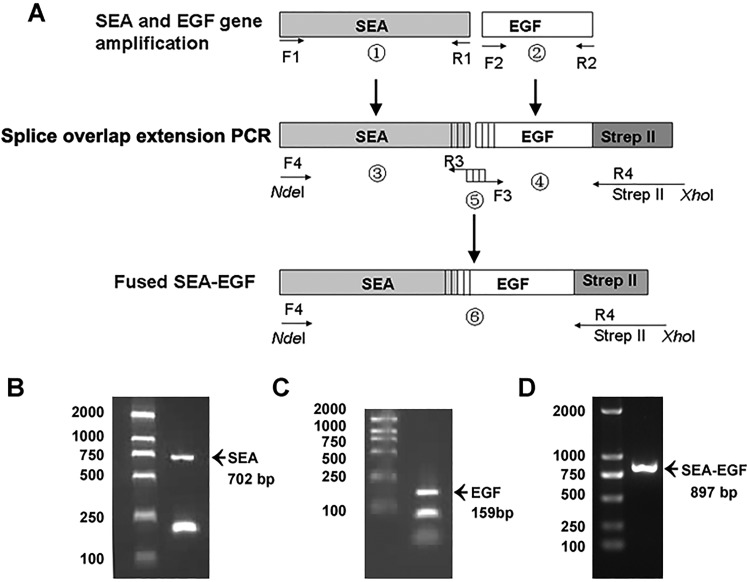
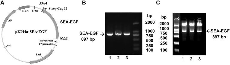
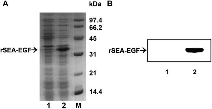
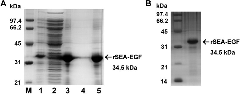
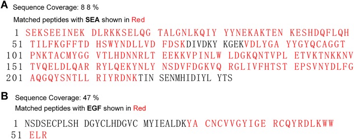
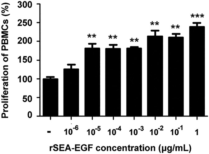
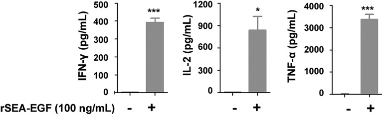
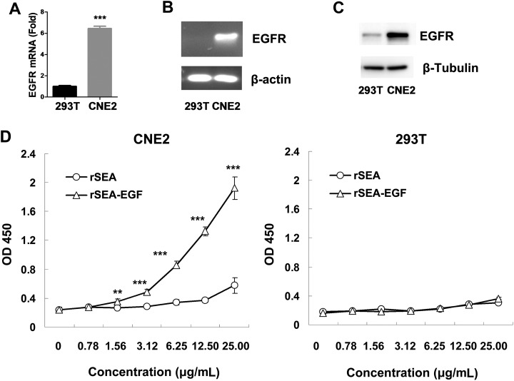
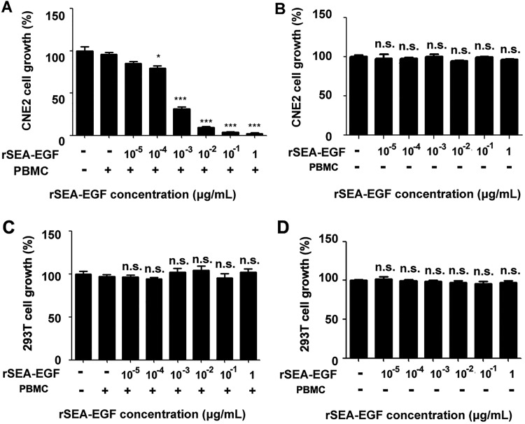
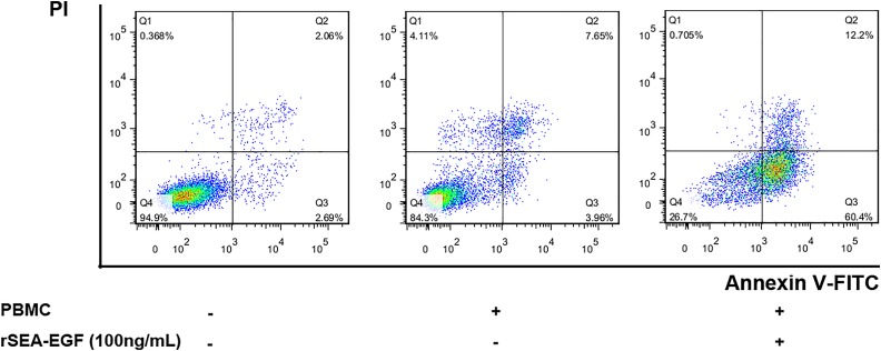
Staphylococcal enterotoxin A is well known as a superantigen and able to be used for cancer immunotherapy. In this study, recombinant Staphylococcal enterotoxin A was genetically conjugated to epidermal growth factor to produce a chimeric protein recombinant Staphylococcal enterotoxin A-epidermal growth factor expressed in Escherichia coli. The recombinant Staphylococcal enterotoxin A-epidermal growth factor protein was purified using Strep-Tactin affinity chromatography and Endotoxin Removal Resin and identified by sodium dodecyl sulfate-polyacrylamide gel electropheresis and liquid chromatography-tandem mass spectrometry analysis. Furthermore, in vitro experiments showed purified recombinant Staphylococcal enterotoxin A-epidermal growth factor could successfully bind to the human nasopharyngeal carcinoma cell line CNE2, significantly promote the proliferation of human peripheral blood mononuclear cells, and enhance the secretion of several cytokines that have broad antitumor activities, such as interferon-γ, tumor necrosis factor-α, and interleukin-2 . Importantly, recombinant Staphylococcal enterotoxin A-epidermal growth factor significantly inhibited proliferation of CNE2 cells and promoted apoptosis in CNE2 cells when cocultured with peripheral blood mononuclear cells. Finally, both the binding of recombinant Staphylococcal enterotoxin A-epidermal growth factor and the toxicity of recombinant Staphylococcal enterotoxin A-epidermal growth factor-activated peripheral blood mononuclear cells were demonstrated as specific and only effective on high epidermal growth factor receptor-expressing cell lines. In all, our work suggests that recombinant Staphylococcal enterotoxin A-epidermal growth factor serves as a promising novel immunotherapeutic agent. More in vivo and in vitro studies are needed to verify its antitumor potency, as well as investigate the underlying mechanisms in cancer immunotherapy.

Keywords: cancer immunotherapy; epidermal growth factor; ligand-targeted therapeutics; nasopharyngeal cancer.; staphylococcal enterotoxin A; superantigen fusion protein.


Conflict of interest statement

Declaration of Conflicting Interests: The author(s) declared no potential conflicts of interest with respect to the research, authorship, and/or publication of this article.

Figures

Figure 1.
Figure 1.
Cloning of Staphylococcal enterotoxin A-epidermal growth factor (SEA-EGF) gene. A, Schematic presentation of splice overlap extension polymerase chain reaction (PCR) for fused SEA-EGF. Epidermal growth factor was fused to the C-terminal of Staphylococcal enterotoxin A (SEA) and tagged with Strep-tag II at the C-terminal. B-D, Polymerase chain reaction amplified products of SEA gene (B), EGF gene (C), and SEA-EGF gene (D), respectively. DNA ladder is indicated on the left side, arrows indicating the PCR products, and their expected sizes are labeled below as well.
Figure 2.
Figure 2.
Identification of the pET44a Staphylococcal enterotoxin A–epidermal growth factor (SEA-EGF) plasmid. A, Plasmid map of the recombinant construct of pET44a-SEA-EGF. B, Identification of the pET44a-SEA-EGF by PCR assay. Lanes 1-3 are agarose gel electrophoresis of PCR products of different clones after using primers for PCR detection. C, Identification of the pET44a-SEA-EGF by restrictive enzyme digestion assay with NdeI and XhoI. Lanes 1-3 are agarose gel electrophoresis of products of different clones after restrictive enzyme digestion.
Figure 3.
Figure 3.
SDS-PAGE and immunoblot analysis of the recombinant Staphylococcal enterotoxin A–epidermal growth factor (rSEA-EGF). A, SDS-PAGE analysis of inducible expression of rSEA-EGF. Lanes 1 and 2: lysates of E coli cells harboring pET44a-SEA before (lane 1) and after (lane 2) IPTG induction. Lane M: Low-molecular-weight standard. B, Western blotting analysis of rSEA-EGF protein with anti-Strep-Tag II antibody. Lanes 1 and 2: Lysates of Escherichia coli cells harboring pET44a SEA-EGF before (lane 1) and after (lane 2) IPTG induction.
Figure 4.
Figure 4.
Identification of refolded and purified recombinant Staphylococcal enterotoxin A-epidermal growth factor (rSEA-EGF) protein. A, SDS-PAGE analysis of inclusion bodies after 2 washes with the wash buffer. Lane M: Low-molecular-weight standard; lane 1: lysates of Escherichia coli cells harboring pET44a Staphylococcal enterotoxin A-epidermal growth factor (SEA-EGF) after IPTG induction. Lane 2, supernatant after cell lysis; Lane 3, pellet after cell lysis (inclusion bodies); Lane 4, supernatant after washing of inclusion bodies; Lane 5, inclusion bodies after washing step. B, SDS-PAGE analysis of the purified rSEA-EGF protein by Strep-Tactin affinity chromatography.
Figure 5.
Figure 5.
Sequence identification of recombinant Staphylococcal enterotoxin A–epidermal growth factor (rSEA-EGF) using liquid chromatography with mass spectrometry (LC-MS/MS) analysis. Amino acid sequence coverage of peptides digested from rSEA-EGF matched with the known SEA (A) and EGF (B) sequences. Matching identified peptides are indicated in bold red.
Figure 6.
Figure 6.
Proliferation of peripheral blood mononuclear cells (PBMCs) stimulated by different concentrations of recombinant Staphylococcal enterotoxin A–epidermal growth factor (rSEA-EGF). Cells were incubated in the presence of different concentrations of rSEA-EGF (1 pg/mL-1 μg/mL) for 7 days at 37°C in 5% CO2. MTS reagent was added for the last 4 hours of the 7-day incubation. **P < .01, ***P < .001, the rSEA-EGF-stimulated human PBMCs compared with the negative control (PBS).
Figure 7.
Figure 7.
Cytokine secretion of peripheral blood mononuclear cells (PBMCs) stimulated by different concentrations of recombinant Staphylococcal enterotoxin A–epidermal growth factor (rSEA-EGF). After 24 hours in culture, the supernatants were harvested and IFN-γ, TNFα, and IL-2 concentration was measured by ELISA assay. The rSEA-EGF-stimulated human PBMCs were compared with the negative control (PBS, *P < .05, ***P < .001) PBMCs. The mean ± standard error of the mean of triplicate determinations is shown.
Figure 8.
Figure 8.
Recombinant Staphylococcal enterotoxin A–epidermal growth factor (rSEA-EGF) binds to epidermal growth factor receptor (EGFR) in CNE2 and HEK 293 T cells. A-C, Expression of EGFR in CNE2 and HEK 293 T cell lines was detected by quantitative PCR (A), RT-PCR assay (B), and Western blot (C). D, The binding of rSEA or rSEA-EGF fusion protein to EGFR. The binding of various concentrations of rSEA or rSEA-EGF fusion proteins to CNE2 cancer cells or HEK 293 T cells was detected by cell ELISA assay. Values represented the mean (standard deviation) of results from 4 samples. **P < .01, ***P < .001, compared with rSEA.
Figure 9.
Figure 9.
Effect of recombinant Staphylococcal enterotoxin A–epidermal growth factor (rSEA-EGF) on proliferation of CNE2 and HEK 293 T cells. A, Peripheral blood mononuclear cells (PBMCs) were treated with rSEA-EGF at different concentrations (10 pg/mL-1 μg/mL) for 7 days. Pretreated PBMCs were then cocultured with CNE2 cells (at a density of 5 × 10³ cells per well) in a total volume of 200 μL in 96-well flat-bottomed plates, at 37°C in 5% CO2 for 72 hours. MTS reagent was added for the last 4 hours of incubation. The mean value of 5 wells was calculated, and each experiment was from at least 1 donor and repeated 3 times. *P< .05, *** P< .001 compared with PBMCs treated with 0 μg/mL rSEA-EGF. B, The same procedure performed as that in Figure 9A but in the absence of PBMCs. C-D, The same procedures performed as that in Figure 9A and 9B on HEK 293 T cells with or without the addition of PBMCs. n.s. indicates nonsignificant.
Figure 10.
Figure 10.
Effect of recombinant Staphylococcal enterotoxin A–epidermal growth factor (rSEA-EGF) on apoptosis in CNE2 cells. Peripheral blood mononuclear cells (PBMCs) were pretreated with rSEA-EGF for 7 days, then added into CNE2 cells and cocultured for 48 hours, followed by annexin V and PI staining and flow cytometry assay. Necrotic cells (Q1, annexin V, PI+), viable cells (Q4, annexin V PI), early (Q3, annexin V+ PI), and late (Q3, annexin V+ PI+) apoptotic cells are defined following the instructions from the Apoptosis Detection Kit (BD Pharmingen), and their percentages are given in the respective regions.

Similar articles

Cited by

References

    1. Chakraborty S, Rahman T. The difficulties in cancer treatment. Ecancermedicalscience. 2012;6:ed16. - PMC - PubMed
    1. Trapani JA, Darcy PK. Immunotherapy of cancer. Aust Fam Physician. 2017;46(4):194–199. - PubMed
    1. Kamta J, Chaar M, Ande A, Altomare DA, Ait-Oudhia S. Advancing cancer therapy with present and emerging immuno-oncology approaches. Front Oncol. 2017;7:64. - PMC - PubMed
    1. Gotwals P, Cameron S, Cipolletta D, et al. Prospects for combining targeted and conventional cancer therapy with immunotherapy. Nat Rev Cancer. 2017;17(5):286–301. - PubMed
    1. Ramagopal UA, Liu W, Garrett-Thomson SC, et al. Structural basis for cancer immunotherapy by the first-in-class checkpoint inhibitor ipilimumab. Proc Natl Acad Sci U S A. 2017;114(21): E4223–E4232. - PMC - PubMed

Publication types

MeSH terms

LinkOut - more resources