Evaluation of skeletal muscle microvascular perfusion of lower extremities by cardiovascular magnetic resonance arterial spin labeling, blood oxygenation level-dependent, and intravoxel incoherent motion techniques
- PMID: 29551091
- PMCID: PMC5858129
- DOI: 10.1186/s12968-018-0441-3
Evaluation of skeletal muscle microvascular perfusion of lower extremities by cardiovascular magnetic resonance arterial spin labeling, blood oxygenation level-dependent, and intravoxel incoherent motion techniques
Abstract
Background: Noninvasive cardiovascular magnetic resonance (CMR) techniques including arterial spin labeling (ASL), blood oxygenation level-dependent (BOLD), and intravoxel incoherent motion (IVIM), are capable of measuring tissue perfusion-related parameters. We sought to evaluate and compare these three CMR techniques in characterizing skeletal muscle perfusion in lower extremities and to investigate their abilities to diagnose and assess the severity of peripheral arterial disease (PAD).
Methods: Fifteen healthy young subjects, 14 patients with PAD, and 10 age-matched healthy old subjects underwent ASL, BOLD, and IVIM CMR perfusion imaging. Healthy young and healthy old participants were subjected to a cuff-induced ischemia experiment with pressures of 20 mmHg and 40 mmHg above systolic pressure during imaging. Perfusion-related metrics, including blood flow, T2* relaxation time, perfusion fraction f, diffusion coefficient D, and pseudodiffusion coefficient D*, were measured in the anterior, lateral, soleus, and gastrocnemius muscle groups. Friedman, Mann-Whitney, Wilcoxon signed rank, and Spearman rank correlation tests were used for statistical analysis.
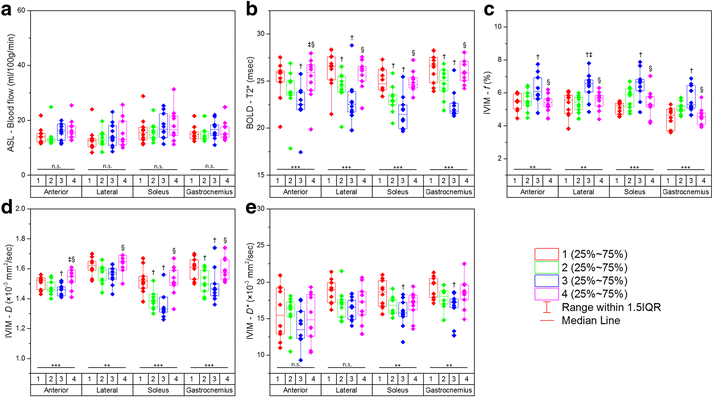
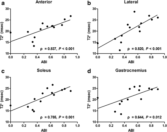
Results: In cases of significant differences determined by the Friedman test (P < 0.05), blood flow, T2*, and D values gradually decreased, while f values showed a tendency to increase in healthy subjects under cuff compression. No significant correlations were found among the ASL, BOLD, and IVIM parameters (all P > 0.05). Blood flow and T2* values showed significant positive correlations with transcutaneous oxygen pressure measurements (ρ = 0.465 and 0.522, respectively; both P ≤ 0.001), while f values showed a significant negative correlation in healthy young subjects (ρ = - 0.351; P = 0.018). T2* was independent of age in every muscle group. T2* values were significantly decreased in PAD patients compared with healthy old subjects and severe PAD patients compared with mild-to-moderate PAD patients (all P < 0.0125). Significant correlations were found between T2* and ankle-brachial index values in all muscle groups in PAD patients (ρ = 0.644-0.837; all P < 0.0125). Other imaging parameters failed to show benefits towards the diagnosis and disease severity evaluation of PAD.
Conclusions: ASL, BOLD, and IVIM provide complementary information regarding tissue perfusion. Compared with ASL and IVIM, BOLD may be a more reliable technique for assessing PAD in the resting state and could thus be applied together with angiography in clinical studies as a tool to comprehensively assess microvascular and macrovascular properties in PAD patients.
Keywords: Blood flow; Cardiovascular magnetic resonance; Diffusion-weighted imaging; Oxygenation; Perfusion.
Conflict of interest statement
Ethics approval and consent to participate
The protocol was approved by the Institutional Review Board at Renji Hospital, School of Medicine, Shanghai Jiao Tong University, and all participants provided written informed consent.
Consent for publication
Not applicable
Competing interests
The authors declare that they have no competing interests.
Publisher’s Note
Springer Nature remains neutral with regard to jurisdictional claims in published maps and institutional affiliations.
Figures


Similar articles
-
Evaluation of skeletal muscle perfusion changes in patients with peripheral artery disease before and after percutaneous transluminal angioplasty using multiparametric MR imaging.Magn Reson Imaging. 2022 Nov;93:157-162. doi: 10.1016/j.mri.2022.08.001. Epub 2022 Aug 5. Magn Reson Imaging. 2022. PMID: 35934206
-
Biexponential analysis of intravoxel incoherent motion in calf muscle before and after exercise: Comparisons with arterial spin labeling perfusion and T2.Magn Reson Imaging. 2020 Oct;72:42-48. doi: 10.1016/j.mri.2020.06.003. Epub 2020 Jun 16. Magn Reson Imaging. 2020. PMID: 32561379
-
Leg blood flow and skeletal muscle microvascular perfusion responses to submaximal exercise in peripheral arterial disease.Am J Physiol Heart Circ Physiol. 2018 Nov 1;315(5):H1425-H1433. doi: 10.1152/ajpheart.00232.2018. Epub 2018 Aug 10. Am J Physiol Heart Circ Physiol. 2018. PMID: 30095999
-
Magnetic Resonance Imaging Techniques in Peripheral Arterial Disease.Adv Wound Care (New Rochelle). 2023 Nov;12(11):611-625. doi: 10.1089/wound.2022.0161. Epub 2023 May 23. Adv Wound Care (New Rochelle). 2023. PMID: 37058352 Free PMC article. Review.
-
Noncontrast Pediatric Brain Perfusion: Arterial Spin Labeling and Intravoxel Incoherent Motion.Magn Reson Imaging Clin N Am. 2021 Nov;29(4):493-513. doi: 10.1016/j.mric.2021.06.002. Magn Reson Imaging Clin N Am. 2021. PMID: 34717841 Review.
Cited by
-
Repeatability of [15O]H2O PET imaging for lower extremity skeletal muscle perfusion: a test-retest study.EJNMMI Res. 2024 Jan 31;14(1):11. doi: 10.1186/s13550-024-01073-x. EJNMMI Res. 2024. PMID: 38294730 Free PMC article.
-
Molecular Imaging of Lower Extremity Peripheral Arterial Disease: An Emerging Field in Nuclear Medicine.Front Med (Lausanne). 2022 Jan 12;8:793975. doi: 10.3389/fmed.2021.793975. eCollection 2021. Front Med (Lausanne). 2022. PMID: 35096884 Free PMC article.
-
Perfusion imaging techniques in lower extremity peripheral arterial disease.Br J Radiol. 2022 Jul 1;95(1135):20211203. doi: 10.1259/bjr.20211203. Epub 2022 May 12. Br J Radiol. 2022. PMID: 35522774 Free PMC article. Review.
-
Advancements in magnetic resonance imaging-based biomarkers for muscular dystrophy.Muscle Nerve. 2019 Oct;60(4):347-360. doi: 10.1002/mus.26497. Epub 2019 May 14. Muscle Nerve. 2019. PMID: 31026060 Free PMC article. Review.
-
Multispectral optoacoustic tomography of peripheral arterial disease based on muscle hemoglobin gradients-a pilot clinical study.Ann Transl Med. 2021 Jan;9(1):36. doi: 10.21037/atm-20-3321. Ann Transl Med. 2021. PMID: 33553329 Free PMC article.
References
-
- Englund EK, Langham MC, Ratcliffe SJ, Fanning MJ, Wehrli FW, Mohler ER, et al. Multiparametric assessment of vascular function in peripheral artery disease dynamic measurement of skeletal muscle perfusion, blood-oxygen-level dependent signal, and venous oxygen saturation. Circ Cardiovasc Imaging. 2015;8:e002673. doi: 10.1161/CIRCIMAGING.114.002673. - DOI - PMC - PubMed
Publication types
MeSH terms
Substances
LinkOut - more resources
Full Text Sources
Other Literature Sources
Medical

