Structure of the Mitochondrial Aminolevulinic Acid Synthase, a Key Heme Biosynthetic Enzyme
- PMID: 29551290
- PMCID: PMC5894356
- DOI: 10.1016/j.str.2018.02.012
Structure of the Mitochondrial Aminolevulinic Acid Synthase, a Key Heme Biosynthetic Enzyme
Abstract
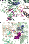
5-Aminolevulinic acid synthase (ALAS) catalyzes the first step in heme biosynthesis. We present the crystal structure of a eukaryotic ALAS from Saccharomyces cerevisiae. In this homodimeric structure, one ALAS subunit contains covalently bound cofactor, pyridoxal 5'-phosphate (PLP), whereas the second is PLP free. Comparison between the subunits reveals PLP-coupled reordering of the active site and of additional regions to achieve the active conformation of the enzyme. The eukaryotic C-terminal extension, a region altered in multiple human disease alleles, wraps around the dimer and contacts active-site-proximal residues. Mutational analysis demonstrates that this C-terminal region that engages the active site is important for ALAS activity. Our discovery of structural elements that change conformation upon PLP binding and of direct contact between the C-terminal extension and the active site thus provides a structural basis for investigation of disruptions in the first step of heme biosynthesis and resulting human disorders.
Keywords: AAA+ unfoldase; ClpX; XLPP; XLSA; porphyria; sideroblastic anemia; α-oxoamine family.
Copyright © 2018 Elsevier Ltd. All rights reserved.
Figures


Similar articles
-
Crystal structure of 5-aminolevulinate synthase, the first enzyme of heme biosynthesis, and its link to XLSA in humans.EMBO J. 2005 Sep 21;24(18):3166-77. doi: 10.1038/sj.emboj.7600792. Epub 2005 Aug 25. EMBO J. 2005. PMID: 16121195 Free PMC article.
-
Crystal structures of aminotransferases Aro8 and Aro9 from Candida albicans and structural insights into their properties.J Struct Biol. 2019 Mar 1;205(3):26-33. doi: 10.1016/j.jsb.2019.02.001. Epub 2019 Feb 8. J Struct Biol. 2019. PMID: 30742897
-
The yeast ALA synthase C-terminus positively controls enzyme structure and function.Protein Sci. 2023 Apr;32(4):e4600. doi: 10.1002/pro.4600. Protein Sci. 2023. PMID: 36807942 Free PMC article.
-
5-Aminolevulinate synthase catalysis: The catcher in heme biosynthesis.Mol Genet Metab. 2019 Nov;128(3):178-189. doi: 10.1016/j.ymgme.2019.06.003. Epub 2019 Jun 13. Mol Genet Metab. 2019. PMID: 31345668 Free PMC article. Review.
-
5-aminolevulinate synthase: catalysis of the first step of heme biosynthesis.Cell Mol Biol (Noisy-le-grand). 2009 Feb 16;55(1):102-10. Cell Mol Biol (Noisy-le-grand). 2009. PMID: 19268008 Free PMC article. Review.
Cited by
-
Influence of Doxorubicin Treatment on Heme Metabolism in Cardiomyoblasts: An In Vitro Study.Arq Bras Cardiol. 2021 Feb;116(2):323-324. doi: 10.36660/abc.20200662. Arq Bras Cardiol. 2021. PMID: 33656083 Free PMC article. English, Portuguese. No abstract available.
-
Regulation of Heme Synthesis by Mitochondrial Homeostasis Proteins.Front Cell Dev Biol. 2022 Jun 27;10:895521. doi: 10.3389/fcell.2022.895521. eCollection 2022. Front Cell Dev Biol. 2022. PMID: 35832791 Free PMC article. Review.
-
Human aminolevulinate synthase structure reveals a eukaryotic-specific autoinhibitory loop regulating substrate binding and product release.Nat Commun. 2020 Jun 4;11(1):2813. doi: 10.1038/s41467-020-16586-x. Nat Commun. 2020. PMID: 32499479 Free PMC article.
-
Recent advances on porphyria genetics: Inheritance, penetrance & molecular heterogeneity, including new modifying/causative genes.Mol Genet Metab. 2019 Nov;128(3):320-331. doi: 10.1016/j.ymgme.2018.11.012. Epub 2018 Nov 30. Mol Genet Metab. 2019. PMID: 30594473 Free PMC article. Review.
-
Mitochondrial ClpX activates an essential biosynthetic enzyme through partial unfolding.Elife. 2020 Feb 24;9:e54387. doi: 10.7554/eLife.54387. Elife. 2020. PMID: 32091391 Free PMC article.
References
-
- Adams PD, Afonine PV, Bunkoczi G, Chen VB, Davis IW, Echols N, Headd JJ, Hung LW, Kapral GJ, Grosse-Kunstleve RW, McCoy AJ, Moriarty NW, Oeffner R, Read RJ, Richardson DC, Richardson JS, Terwilliger TC, Zwart PH. PHENIX: a comprehensive Python-based system for macromolecular structure solution. Acta crystallographica. Section D, Biological crystallography. 2010;66:213–221. - PMC - PubMed
-
- Balwani M, Bloomer J, Desnick R. X-Linked Protoporphyria. In: Pagon RA, Adam MP, Ardinger HH, Wallace SE, Amemiya A, Bean LJH, Bird TD, Ledbetter N, Mefford HC, Smith RJH, Stephens K, editors. GeneReviews(R) Seattle (WA): 1993. - PubMed
-
- Balwani M, Doheny D, Bishop DF, Nazarenko I, Yasuda M, Dailey HA, Anderson KE, Bissell DM, Bloomer J, Bonkovsky HL, Phillips JD, Liu L, Desnick RJ. Loss-of-function ferrochelatase and gain-of-function erythroid-specific 5-aminolevulinate synthase mutations causing erythropoietic protoporphyria and x-linked protoporphyria in North American patients reveal novel mutations and a high prevalence of X-linked protoporphyria. Mol Med. 2013;19:26–35. - PMC - PubMed
Publication types
MeSH terms
Substances
Grants and funding
LinkOut - more resources
Full Text Sources
Other Literature Sources
Molecular Biology Databases
Research Materials

