Review
. 2018 Oct:368:28-40.
doi: 10.1016/j.heares.2018.03.002. Epub 2018 Mar 11.

Pharmacokinetic principles in the inner ear: Influence of drug properties on intratympanic applications

Affiliations
Review

Pharmacokinetic principles in the inner ear: Influence of drug properties on intratympanic applications

Alec N Salt et al. Hear Res. 2018 Oct.

Abstract

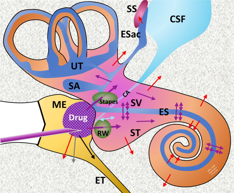
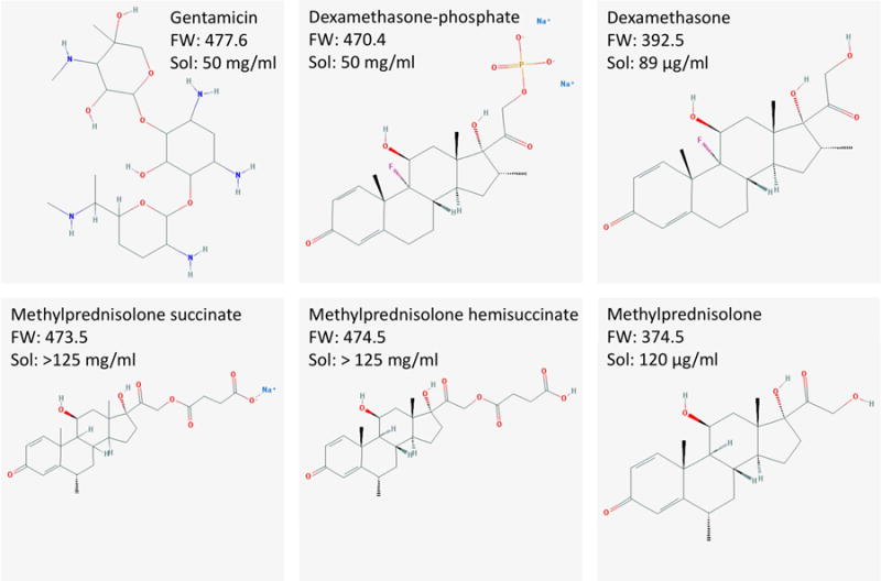
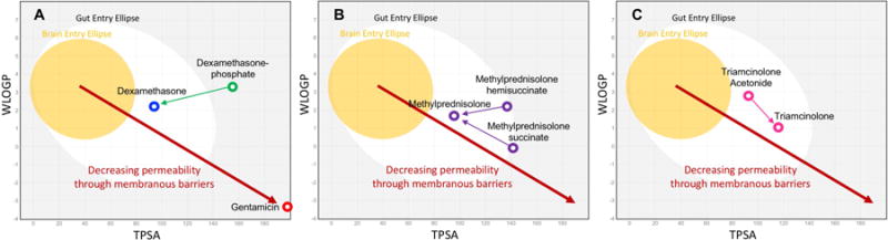
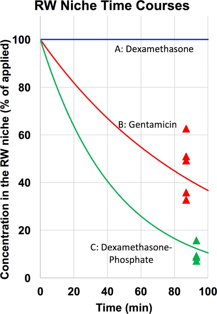
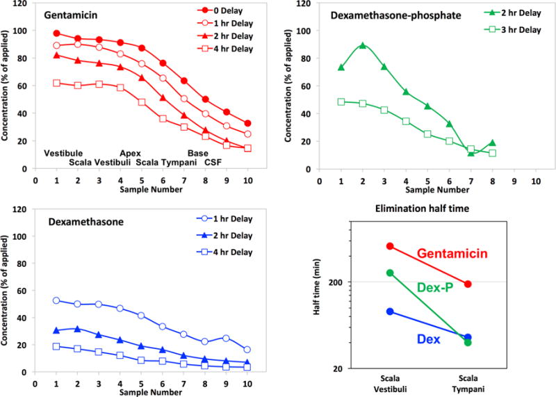
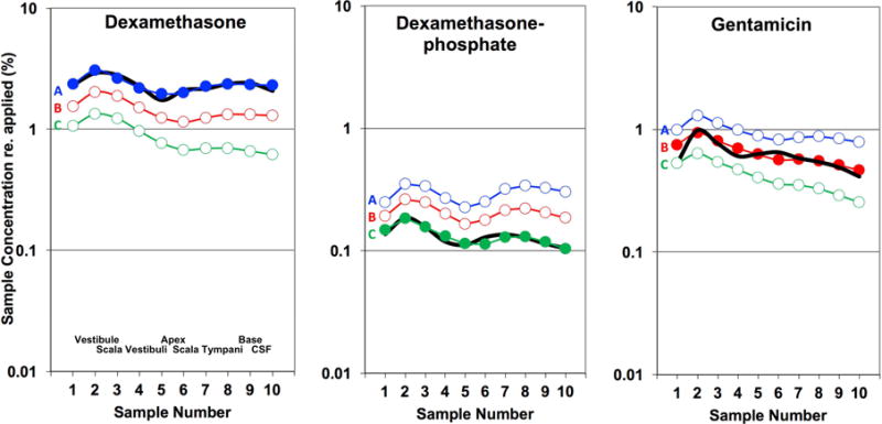
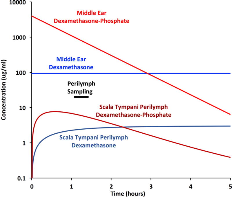
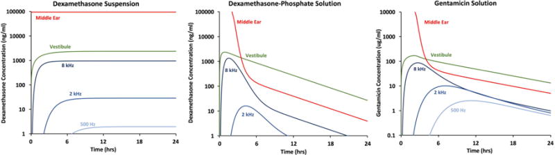
Local drug delivery to the ear has gained wide clinical acceptance, with the choice of drug and application protocol in humans largely empirically-derived. Here, we review the pharmacokinetics underlying local therapy of the ear using the drugs commonly used in clinical practice as examples. Based on molecular properties and perilymph measurements interpreted through computer simulations we now better understand the principles underlying entry and distribution of these and other drugs in the ear. From our analysis, we have determined that dexamethasone-phosphate, a pro-drug widely-used clinically, has molecular and pharmacokinetic properties that make it ill-suited for use as a local therapy for hearing disorders. This polar form of dexamethasone, used as a more soluble agent in intravenous preparations, passes less readily through lipid membranes, such as those of the epithelia restricting entry at the round window membrane and stapes. Once within the inner ear, dexamethasone-phosphate is cleaved to the active form, dexamethasone, which is less polar, passes more readily through lipid membranes of the blood-perilymph barrier and is rapidly eliminated from perilymph without distributing to apical cochlear regions. Dexamethasone-phosphate therefore provides only a brief exposure of the basal regions of the cochlea to active drug. Other steroids, such as triamcinolone-acetonide, exhibit pharmacokinetic properties more appropriate to the ear and merit more detailed consideration.

Keywords: Drug delivery; Inner ear; Intracochlear; Intralabyrinthine; Oval window; Perilymph; Round window membrane.


Figures

Figure 1
Figure 1
Schematic of drug applied intratympanically to the inner ear. Colored arrows indicate movements of drug; Purple: Distribution; Red: Elimination to blood; Cyan: CSF-Perilymph fluid exchange; Gray: Elimination to lymphatics; Black: Elimination via the Eustachian tube. Abbreviations are: CSF: Cerebrospinal Fluid; CA: Cochlear aqueduct; Esac: Endolymphatic Sac; ES: Endolymphatic Space; ET: Eustachian tube; ME: Middle Ear; RW: Round Window; SA: Saccule; SS: Sigmoid Sinus; ST: Scala Tympani; SV: Scala Vestibuli; UT: Utricle.
Figure 2
Figure 2
Drugs in common use for intratympanic therapy of the inner ear. Formula weights (FW) given do not include counter ions from which they dissociate in solution. “Sol” indicates the solubility in aqueous solution. Formulae courtesy of the Pubchem website (https://pubchem.ncbi.nlm.nih.gov).
Figure 3
Figure 3
Membrane permeation-related characteristics, WLOGP (lipid solubility, Y-axis) and TPSA (topological polar surface area; X-axis) for a number of drugs calculated by the SwissADME website (http://http://www.swissadme.ch). The yellow ellipse bounds the statistical range for molecules that pass through the blood-brain barrier and the white ellipse bounds the range for molecules that permeate the gut (Daina & Zoete, 2016; Daina et al., 2017). Based on this analysis, dexamethasone and methylprednisolone would be expected to permeate membranes more readily than dexamethasone-phosphate, methylprednisolone-hemisuccinate, and methylprednisolone-succinate. Gentamicin would be expected to be substantially less permeable than all forms of the steroids.
Figure 4
Figure 4
Data points show the measured drug concentration remaining in the RW niche in perilymph sampling experiments that used a single 20 μL application to the RW niche in the guinea pig. For dexamethasone-phosphate the measure was the sum of that metabolized to the base form (72%) and that remaining as the phosphate form (28%). Subsequent calculations have assumed an exponential decline of concentration with time as indicated by the curves. For dexamethasone applied as a suspension of micronized solid drug, a uniform concentration over the application period was assumed in calculations.
Figure 5
Figure 5
Perilymph elimination compared by perilymph loading followed by sequential sampling from the LSCC after the delay times indicated. The approximate origins of the samples are indicated in the first panel. These curves show the progressive decline of perilymph drug concentration with time. Dexamethasone and dexamethasone-phosphate data are from Salt et al., (2012b) and gentamicin data are from Salt et al., (2016).
Figure 6
Figure 6
Perilymph concentrations resulting from RW niche applications for 3 drugs. Black lines show the group mean measured perilymph concentrations in 10 sequential samples taken from the lateral semi-circular canal. The number of experiments were 4, 4 and 5 respectively. Concentrations are normalized (as %) of the drug concentration applied to the RW niche. The applied concentration of dexamethasone was 94.2 μg/ml (dissolved concentration in an applied 4.5% suspension), of dexamethasone-phosphate was 4 mg/ml and of gentamicin was 40 mg/ml respectively. The approximate spatial origins of the perilymph samples are given in the first panel. Colored curves are simulations of the experiments calculated for each of the 3 time courses of drug decline in the middle ear. A, blue: unchanging with time, comparable to micronized dexamethasone suspension, B, red: 74.5 min middle ear decline half-time, comparable to gentamicin solution, C: green: 25.7 min middle ear decline half-time, comparable to dexamethasone phosphate. These decline rates correspond to the curves shown in Figure 4, and are detailed in the text. Dexamethasone and dexamethasone-phosphate data are from Salt et al., 2018 and gentamicin data are from Salt et al., 2016.
Figure 7
Figure 7
Time courses of dexamethasone-phosphate and dexamethasone base concentrations derived from simulations fitted to the measured sample data. Concentrations are shown for perilymph of the entire scala tympani pooled. Perilymph concentration is initially higher with application of dexamethasone-phosphate, due to the higher applied concentration, and was higher at the time of sampling which commenced after 1 hour as indicated. Nevertheless, the higher dosing is short-lived and after 2.3 hours concentration falls below that when dexamethasone suspension is administered.
Figure 8
Figure 8
Influence of drug properties and application protocols on calculated drug levels achieved in different regions of the human inner ear using kinetic parameters established in the studies above. Sustained delivery from a suspension of dexamethasone results in the generation of a steady-state, with substantial concentration gradients between the vestibule and the cochlea, and between high-frequency and low-frequency cochlear regions. In contrast, delivery of dexamethasone-phosphate solution results in a transient drug exposure, primarily to vestibular and high-frequency cochlear regions, due to the rapid rate of elimination of dexamethasone from perilymph. Delivery of gentamicin also results in a transient drug exposure, with greater spread to low frequency cochlear regions then dexamethasone-phosphate, due to a lower rate of elimination from cochlear perilymph.

Similar articles

Cited by

References

    1. Alexander TH, Harris JP, Nguyen QT, Vorasubin N. Dose Effect of Intratympanic Dexamethasone for Idiopathic Sudden Sensorineural Hearing Loss: 24 mg/mL Is Superior to 10 mg/mL. Otol Neurotol. 2015;36:1321–1327. - PubMed
    1. Ayoob AM, Borenstein JT. The role of intracochlear drug delivery devices in the management of inner ear disease. Expert Opin Drug Deliv. 2015;12:465–479. - PubMed
    1. Bird PA, Begg EJ, Zhang M, Keast AT, Murray DP, Balkany TJ. Intratympanic versus intravenous delivery of methylprednisolone to cochlear perilymph. Otol Neurotol. 2007;28:1124–1130. - PubMed
    1. Briggs R, O’leary S, Birman C, et al. A first time in human investigation of a combination device delivering a targeted drug therapy to cochlear implant recipients. 14th International Conference on Cochlear Implants; Toronto. 2016.
    1. Chen Z, Kujawa SG, McKenna MJ, Fiering JO, Mescher MJ, Borenstein JT, Swan EE, Sewell WF. Inner ear drug delivery via a reciprocating perfusion system in the guinea pig. J Control Release. 2005;110:1–19. - PMC - PubMed

Publication types

Substances