. 2018 Jul:172:10-20.
doi: 10.1016/j.exer.2018.03.015. Epub 2018 Mar 15.

Expression and regulation of alarmin cytokine IL-1α in human retinal pigment epithelial cells

Affiliations

Expression and regulation of alarmin cytokine IL-1α in human retinal pigment epithelial cells

Zong-Mei Bian et al. Exp Eye Res. 2018 Jul.

Abstract

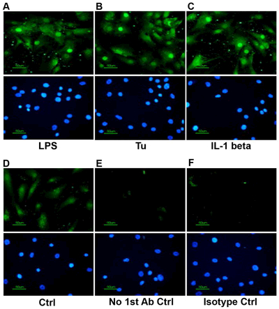
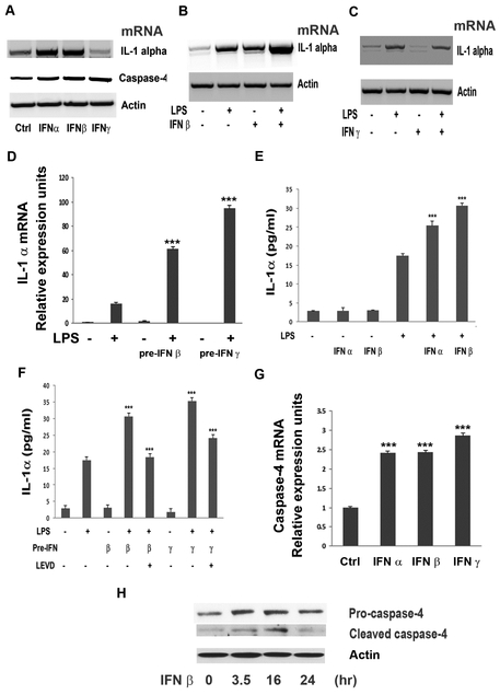
Human retinal pigment epithelial (hRPE) cells play important immune-regulatory roles in a variety of retinal pathologic processes, including the production of inflammatory cytokines that are essential mediators of the innate immune response within the ocular microenvironment. The pro-inflammatory "alarmin" cytokine IL-1α has been implicated in both infectious and non-infectious retinal diseases, but its regulation in the retina is poorly understood. The purpose of this study was to elucidate the expression and regulation of IL-1α within hRPE cells. To do this, IL-1α mRNA and protein in hRPE cells was assessed by RT-PCR, qPCR, ELISA, Western blot, and immunofluorescence following treatment with a variety of stimuli and inhibitors. ER stress, LPS, IL-1β, and TLR2 activation all significantly increased intracellular IL-1α protein. Increasing intracellular calcium synergized both LPS- and Pam3CSK4-induced IL-1α protein production. Accordingly, blocking calcium signaling and calpain activity strongly suppressed IL-1α protein expression. Significant but more moderate inhibition occurred following blockage of TLR4, caspase-4, or caspase-1. Neutralizing antibodies to IL-1β and TLR2 partially eliminated LPS- and TLR2 ligand Pam3CSK4-stimulated IL-1α protein production. IFN-β induced caspase-4 expression and activation, and also potentiated LPS-induced IL-1α expression, but IFN-β alone had no effect on IL-1α protein production. Interestingly, all inhibitors targeting the PI3K/Akt pathway, with the exception of Ly294002, strongly increased IL-1α protein expression. This study improves understanding of the complex mechanisms regulating IL-1α protein expression in hRPE cells by demonstrating that TLR4 and TLR2 stimulation and exposure to IL-1β, ER stress and intracellular calcium all induce hRPE cells to produce intracellular IL-1α, which is negatively regulated by the PI3K/Akt pathway. Additionally, the non-canonical inflammasome pathway was shown to be involved in LPS-induced hRPE IL-1α expression through caspase-4 signaling.

Keywords: Caspase-4; IL-1α; Interleukin-1α; Non-canonical inflammasome; RPE; Retinal pigment epithelium; TLR2; TLR4.


Figures

Fig 1.
Fig 1.
IL-1α mRNA synthesis and protein production in hRPE cells. mRNA levels were assessed for cells (A) treated with LPS (1000 ng/ml), tunicamycin (10 μM) or IL-1β (2 ng/ml) for 6 hr. IL-1α protein production was analyzed by ELISA in hRPE cells treated with LPS (B, 1000 ng/ml), tunicamycin (C, 10 μM) or IL-1β (D, 2 ng/ml) for 24 or 48 hr in the presence or absence of caspase-4 inhibitor (LEVD, 4 μM), caspase-1 inhibitor (YVAD, 2 μM), anti-IL-1β antibody (Ab IL-1β) or isotype serium (Ab Ctrl). The p values were calculated by comparing treatment to Ctrl (A), to LPS only (B), to tunicamycin only (C), or to IL-1β only (D). *p<0.05; **p<0.01; ***p<0.001. In order to compare IL-1α mRNA levels under different conditions, expression of the house keeping gene β-action was used to monitor gel loading. Ab, antibody; Ctrl, control; LEVD, Ac-LEVD-CHO; YVAD, Ac-YVAD-cmk.
Fig. 2.
Fig. 2.
Immunofluorescence analysis of IL-1α expression in hRPE cells. hRPE cells seeded in chamber slides were incubated with LPS (A, 1000 ng/ml), tunicamycin (B, 10 μM) or IL-1β (C, E, F, 2 ng/ml) for 20 hr. Controls include untreated cells (D), IL-1β-treated cells without primary antibody (E), and IL-1β-treated cells with an isotype antibody control (F). The cells were fixed as described in Materials and Methods. The expressed IL-1α is shown in green. The nuclei were stained with 400x bisBenzimide (blue) (A-F, bottom panels). Experiments were repeated twice. Typical images were taken at 400X magnification.
Fig. 3.
Fig. 3.
Effects of IFNs on caspase-4 expression/activation and LPS-induced IL-1α production. mRNA levels of IL-1α and caspase-4 were analyzed by RT-PCR (A-C) and qPCR (D, G). IL-1α protein was analyzed by ELISA (E and F). Caspase-4 protein cleavage in hRPE cells was shown by Western blot analysis (H). hRPE cells were treated with LPS (1000 ng/ml), IFN-α (1000 U/ml), IFN-β (1000 U/ml), IFN-γ (1000 U/ml), IFN-γ priming or in combination for 6 (A-D, G), 24 (E-F) or 0, 16 and 24 hr (H). In E and F, hRPE cells were pre-incubated with IFN-α, -β and -γ at 1000 U/ml for 16 hr and then switched to fresh growth media with or without LPS (1000 ng/ml) for an additional 24 hr. The p values were calculated by comparing treatment with LPS alone (D, E, F), treatment with LPS plus caspase-4 inhibitor (F), or treatment with control (G). *p<0.05; **p<0.01; ***p<0.001. Ctrl, control; IFN, interferon; LEVD, Ac-LEVD-CHO; pre-, pre-incubation with.
Fig. 4.
Fig. 4.
Modulation of IL-1α expression through TLR4 and TLR2 signaling. hRPE cells were cultured with LPS (1000 ng/ml) (A, B) or Pam3CSK4 (Pam 300 ng/ml) or LPS plus 100 ng/ml Pam3CSK4 (B, C) for 24 hr. In some experiments, hRPE cells also were pre-incubated with TAK-242 (75 μM) (A), caspase-4 inhibitor LEVD-CHO (4 μM) (C), TLR2 blocking antibody, or isotype control antibody (C). After stimulations, whole cell lysates were harvested and subjected to ELISA. *p<0.05; **p<0.01; ***p<0.001, as compared with inducers alone. Ab T2, anti-TLR2 antibody; Ab Ctrl, isotype control; LEVD, LEVD-CHO; Pam, Pam3CSK4; TAK, TAK-242.
Fig. 5.
Fig. 5.
Ca2+ signaling regulates IL-1α expression. hRPE cells were cultured with or without LPS (1000 ng/ml) (A and F), tunicamycin (Tu, 10μM) (B), IL-1β (2 ng/ml) (C), or Pam3CSK4 (Pam, 100 ng/ml) (D) for 6 (F) or 24 hr (A-D) with or without PD150606 (PD, 100 μM) or BAPTA-AM (BAPTA, 5 μM). hRPE cells were incubated with or without ionomycin (3 μM), LPS, and Pam3CSK4 in presence or absence of caspase-4 inhibitor, LEVD (4 μM), and inhabited with ionomycin plus LPS or ionomycin plus Pam3CSK4 for 24 hr (E). After incubation, the whole cell lysates were harvested and subjected to ELISA. Steady-state IL-1α mRNA was analyzed by qPCR (F). *p<0.05; **p<0.01; ***p<0.001, as compared with inducers alone (A-E), or with control (B).
Fig. 6.
Fig. 6.
The effect of CU-CPT22 on IL-1α expression. hRPE cells were cultured with or without CU-CPT22 or co-cultured with LPS (1000 ng/ml) or Pam3CSK4 (100 ng/ml) for 24 hr (A-C). Unless specified, the concentration of CU-CPT22 was 10 μM, TAK-242 100μM, BAPTA-AM 5 μM, caspase-4 inhibitor, LEVD 4 μM, Ly 294002 75 μM. After incubation, the whole cell lysates were harvested and subjected to ELISA. *p<0.05; **p<0.01; ***p<0.001, as compared with inducers alone (A-C) or control (A, B). Ab T2 anti-TLR2 antibody; Ab Ctrl, isotype control; CU, CU-CPT22; BAPTA, BAPTA-AM; LEVD, LEVD-CHO; Ly, Ly 294002; Pam, Pam3CSK4; TAK, TAK-242.
Fig. 7.
Fig. 7.
Effects of PI3K inhibitors on IL-1α expression. hRPE cells were cultured with or without LPS (1000 ng/ml) in the presence or absence of CAL-101, PI-103, Wortmannin and MK 2206 for 6 hr to measure mRNA levels (A, C) or 24 hr to measure protein (B, D). ***p<0.001, as compared with inducers alone.
Fig. 8.
Fig. 8.
A schematic view of pathways regulating IL-1α expression in hRPE cells. LPS induced IL-1α expression is mediated by two signal pathways: the TLR4 receptor pathway and the non-canonical inflammasome pathway. After stimulation with LPS, TLR4 binds the sorting adaptor TIRAP, forming an early-acting TIRAP/MyD88 complex, which activates NF-kB, leading to IL-1α expression. An alternative pathway, also involving TIRAP/MyD88, appears to mediate pro-IL-1α expression through TRL2 binding by its agonist, PAM3CSK4. However, the pro-inflammatory response by LPS is subject to negative regulation by PI3K, which stimulates dissociation and degradation of TIRAP and triggers a late-acting anti-inflammatory phase (dotted line), in which TLR4 recruits the TRIF/TRAM complex and induces endosomal-internalization. In this process, LPS may also to be internalized. The intracellular [LPS]in, as with ER-stress and IFNs, mediates non-canonical activation of the inflammasome leading to production of IL-1α. The roles of calcium-dependent calpain in IL-1α production may be due to its stimulation of IκBα degradation, which promotes pro-IL-1α maturation. The inflammasome-mediated release of IL-1β further induces IL-1α expression via an autocrine mechanism. The numbers represent corresponding references: 1 (Aksoy et al., 2012; Siegemund and Sauer, 2012); 2 (Husebye et al., 2006); 3 (Shi et al., 2014); 4 (Bian et al., 2009); 5 (Kayagaki et al., 2015); 6 (Schaecher et al., 2004); 7 (Kavita and Mizel, 1995); 8 (Cui et al., 2013).

Similar articles

Cited by

References

    1. Agrawal S, Gupta S, 2011. TLR1/2, TLR7, and TLR9 signals directly activate human peripheral blood naive and memory B cell subsets to produce cytokines, chemokines, and hematopoietic growth factors. J Clin Immunol 31, 89–98. - PMC - PubMed
    1. Aksoy E, Taboubi S, Torres D, Delbauve S, Hachani A, Whitehead MA, Pearce WP, Berenjeno IM, Nock G, Filloux A, Beyaert R, Flamand V, Vanhaesebroeck B, 2012. The p110delta isoform of the kinase PI(3)K controls the subcellular compartmentalization of TLR4 signaling and protects from endotoxic shock. Nat Immunol 13, 1045–1054. - PMC - PubMed
    1. Bandyopadhaya A, Bhowmick S, Chaudhuri K, 2009. Activation of proinflammatory response in human intestinal epithelial cells following Vibrio cholerae infection through PI3K/Akt pathway. Can J Microbiol 55, 1310–1318. - PubMed
    1. Bian ZM, Elner SG, Elner VM, 2009. Dual involvement of caspase-4 in inflammatory and ER stress-induced apoptotic responses in human retinal pigment epithelial cells. Investigative ophthalmology & visual science 50, 6006–6014. - PMC - PubMed
    1. Bian ZM, Elner SG, Yoshida A, Elner VM, 2003. Human RPE-monocyte co-culture induces chemokine gene expression through activation of MAPK and NIK cascade. Experimental eye research 76, 573–583. - PubMed

Publication types

MeSH terms