. 2018 Jun:46:154-165.
doi: 10.1016/j.cellsig.2018.03.006. Epub 2018 Mar 15.

Dictyostelium Erk2 is an atypical MAPK required for chemotaxis

Affiliations

Dictyostelium Erk2 is an atypical MAPK required for chemotaxis

David J Schwebs et al. Cell Signal. 2018 Jun.

Abstract

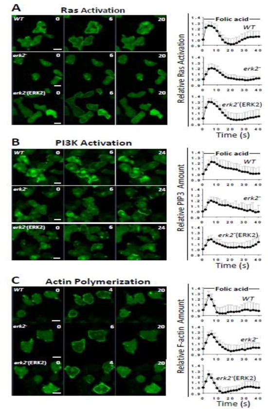
The Dictyostelium genome encodes only two MAPKs, Erk1 and Erk2, and both are expressed during growth and development. Reduced levels of Erk2 expression have been shown previously to restrict cAMP production during development but still allow for chemotactic movement. In this study the erk2 gene was disrupted to eliminate Erk2 function. The absence of Erk2 resulted in a complete loss of folate and cAMP chemotaxis suggesting that this MAPK plays an integral role in the signaling mechanisms involved with this cellular response. However, folate stimulation of early chemotactic responses, such as Ras and PI3K activation and rapid actin filament formation, were not affected by the loss of Erk2 function. The erk2- cells had a severe defect in growth on bacterial lawns but assays of bacterial cell engulfment displayed only subtle changes in the rate of bacterial engulfment. Only cells with no MAPK function, erk1-erk2- double mutants, displayed a severe proliferation defect in axenic medium. Loss of Erk2 impaired the phosphorylation of Erk1 in secondary responses to folate stimulation indicating that Erk2 has a role in the regulation of Erk1 activation during chemotaxis. Loss of the only known Dictyostelium MAPK kinase, MekA, prevented the phosphorylation of Erk1 but not Erk2 in response to folate and cAMP confirming that Erk2 is not regulated by a conventional MAP2K. This lack of MAP2K phosphorylation of Erk2 and the sequence similarity of Erk2 to mammalian MAPK15 (Erk8) suggest that the Dictyostelium Erk2 belongs to a group of atypical MAPKs. MAPK activation has been observed in chemotactic responses in a wide range of organisms but this study demonstrates an essential role for MAPK function in chemotactic movement. This study also confirms that MAPKs provide critical contributions to cell proliferation.

Keywords: Chemotaxis; Dictyostelium; Erk1; Erk2; MAPK.


Figures

Figure 1
Figure 1. Disruption and knock-in complementation of the erk2 locus
A) Homologous recombination of the erk2thyA fragment (see Materials and Methods for construction) with the erk2 locus. The location of primer binding sites (arrows) used for PCR verification of recombination are shown. Open rectangles represent the erk2 open reading frame, the closed rectangle represents the open reading from of an adjacent gene and the thick black line represents the thyA genomic fragment. B) Knock-in of an Erk2 expression vector with blasticidin resistance into the disrupted erk2thyA locus at the SphI site. Hashed lines represent sequences not shown to reduce the size of image. Description of PCR products and primer sequences are described in Fig. S1. C) Immunoblot of Erk2 protein in wild-type (WT), erk2, and erk1erk2 strains and in mutant strains complemented with Erk2 expression vector (Erk2). Lysates of cells grown in axenic medium were analyzed for Erk2 protein by immunoblot analysis. Coomassie staining of the gel was used as a lane loading control.
Figure 2
Figure 2. Dictyostelium growth
A) Growth of MAPK mutants and wild-type cells on bacterial lawns. Individual strains were mixed with bacteria and plated on SM+/3 plates as described in the Materials and Methods section. Images of plaques were captured 5 days later. All images are the same magnification. B) Growth of MAPK mutants and wild-type cells in shaking cultures of axenic medium. Wild-type (WT), erk2, erk1erk2, and the erk2 mutant strains complemented Erk2 expression vectors (Erk2) were inoculated into shaking cultures of HL-5 axenic medium and cells concentrations were determined using a hemacytometer at the indicated times. Each data point represents 4 counts of at least 100 cells. Error bars represent standard deviation in multiple counts.
Figure 3
Figure 3. Engulfment of bacteria
A) Wild-type (WT), far1, and erk2 strains and erk2 mutant complemented with Erk2 expression vector (Erk2) were mixed with pHrodo-labelled live bacteria and analyzed at indicated times for the percentage of pHrodo-positive cells. B) Graphical representation of data from (A). C) Images of engulfed bacteria in cells after 15 min. D) Quantitation of bacterial cell uptake into cells. The engulfed bacterial number in each cell was measured and plotted.
Figure 4
Figure 4. Chemotaxis of MAPK mutants to folate
A) Above-agar chemotaxis assay images for wild-type (WT), erk2, and erk1erk2 strains and erk2 mutants complemented with Erk2 vector (Erk2) after 2.5 h exposure to droplets of 100 μM folate. A) Relative movement of wild-type (WT), erk2, erk1erk2, gα4 and far1 strains and MAPK mutants complemented with Erk2 expression vector (Erk2) toward folate (filled bars) and relative movement in the absence of folate (open bars). Values indicate maximum distance of cell migration toward the source of folate or migration in any direction in the absence of folate. Error bars represent the standard deviation of the error. B) Cell migration paths of select cells were mapped over a 30 min period using time-lapse photography as described in the Materials and Methods section. All images are the same magnification. C) Graphical representation of the average path lengths in arbitrary units (a.u.) from (B). Error bars represent standard deviation. Student’s unpaired t-test comparing to WT, P<0.0001 (*).
Figure 5
Figure 5. Early chemotactic signaling in response to folate
Translocation of Ras, PI3K, and actin filament reporters in wild-type (WT), erk2 cells (erk2), and complemented erk2 cells (Erk2) in response to folate stimulation was assayed as described in the Materials and Methods section. A) Translocation of the Ras activation reporter RBD-GFP to the membrane. B) Translocation of the PI3K activation reporter PHCRAC-GFP to the membrane. C) Translocation of the actin filament reporter LimEΔcoil-GFP to the membrane. Graphs indicate relative intensity of fluorescence at the membrane and 1 represents the intensity at the start of the response. Error bars represent standard deviation. All images are the same magnification and scale bar represents 5 μm.
Figure 6
Figure 6. Development and cAMP chemotaxis
A) Wild-type (WT), erk2, and erk1-erk2 mutants and mutants complemented with Erk2 expression vector (Erk2) developed on nonnutrient plates for 13 h. All images are the same magnification. B) A GFP vector was used to label erk2, erk1erk2, and wild-type (WT) cells. Labeled cells (GFP) were mixed in a 1:9 ratio with unlabeled wild-type cells and and cell droplets (1×10⁷ cells/ml) plated for development on nonnutrient agar plates. Images of aggregation streams were taken at 12 h. All images are the same magnification. C) Above-agar cAMP chemotaxis assay. After 4 h of starvation in shaking phosphate buffer cells were plated on nonnutrient plates near droplets of 100 μM cAMP. Images of cells were taken at 0 h and 2.5 h and distance was measured of the leading edge of cells toward the source of cAMP. Migration distances under 100 μm are typical for random movement in the absence of exogenous cAMP. Error bars represent the standard deviation of the error. Student’s unpaired t-test comparing to WT, P<0.0001 (*).
Figure 7
Figure 7. Phosphorylation of MAPKs
A) After 50 μM folate stimulation erk2 and wild-type (WT) were lysed at times indicated and analyzed for the phosphorylation of MAPKs by immunoblots using phospho-MAPK specific antibodies (upper panel). Coomassie blue stained gel as loading control (lower panel). B) Phosphorylation of MAPKs in mekA cells in response to folate or cAMP. Cells were stimulated with either 50 μM folate or 100 nM cAMP and then analyzed for phosphorylation of the MAPKs as described in (A) (upper panel). Detection of CCCM using HRP-streptavidin as a loading control (lower panel).
Figure 8
Figure 8. Phylogenetic analysis of MAPKs
All known MAPKs in human (Hs), yeast/Saccharomyces cerevisiae (Sc), and Dictyostelium discoideum (Dd) were used to construct the phylogenetic tree using MEGA7 as described in the Materials and Methods. Selected MAPKs with similarity to atypical human MAPK15 (Erk8) from Drosophila melanogaster (Dm) and Acanthamoeba castellanii (Ac) were also included in the tree. A BLAST search of fungal genomes using the Dictyostelium Erk2 protein as the query yielded only MAPKs with similarities to the human Erk1/2 group such as the one representative MAPK included from Aspergillus nidulans (An).
Figure 9
Figure 9. Model of Erk2 mediated signaling pathways
Multiple chemoattractant stimulated pathways lead to the activation of Erk2 and downstream cellular responses such as chemotaxis and Erk1 activation. Early chemotactic responses such as Ras and PI3K activation and actin filament formation are not dependent on Erk2 function. Like mammalian atypical MAPKs, the activation of Erk2 does not require the only known MAP2K in Dictyostelium. Folate but not cAMP responses require G protein function for Erk2 activation.

Similar articles

Cited by

References

    1. Meloche S, Pouyssegur J. The ERK1/2 mitogen-activated protein kinase pathway as a master regulator of the G1- to S-phase transition. Oncogene. 2007;26(22):3227–39. - PubMed
    1. Pearson G, Robinson F, Beers Gibson T, Xu BE, Karandikar M, Berman K, Cobb MH. Mitogen-activated protein (MAP) kinase pathways: regulation and physiological functions. Endocr Rev. 2001;22(2):153–83. - PubMed
    1. Hadwiger JA, Nguyen HN. MAPKs in development: insights from Dictyostelium signaling pathways. Biomol Concepts. 2011;2(1–2):39–46. - PMC - PubMed
    1. Chen RE, Thorner J. Function and regulation in MAPK signaling pathways: lessons learned from the yeast Saccharomyces cerevisiae. Biochimica et biophysica acta. 2007;1773(8):1311–40. - PMC - PubMed
    1. Cargnello M, Roux PP. Activation and function of the MAPKs and their substrates, the MAPK-activated protein kinases. Microbiology and molecular biology reviews: MMBR. 2011;75(1):50–83. - PMC - PubMed

Publication types

MeSH terms

LinkOut - more resources