

Blue Nevi of the Ocular Surface: Clinical Characteristics, Pathologic Features, and Clinical Course
- PMID: 29551421
- PMCID: PMC6056321
- DOI: 10.1016/j.ophtha.2018.02.006
Blue Nevi of the Ocular Surface: Clinical Characteristics, Pathologic Features, and Clinical Course
Abstract
Purpose: Blue nevus is a melanocytic tumor that is commonly found in the skin. Extracutaneous presentations, including the ocular surface, are rare. As such, the purpose of this study was to characterize the clinical features and clinical course of congenital melanocytic tumor (blue nevus) of the conjunctiva.
Design: Retrospective, noncomparative case series.
Participants: Twenty-one patients with 23 blue nevi of the ocular surface that were excised surgically between 2000 and 2016.
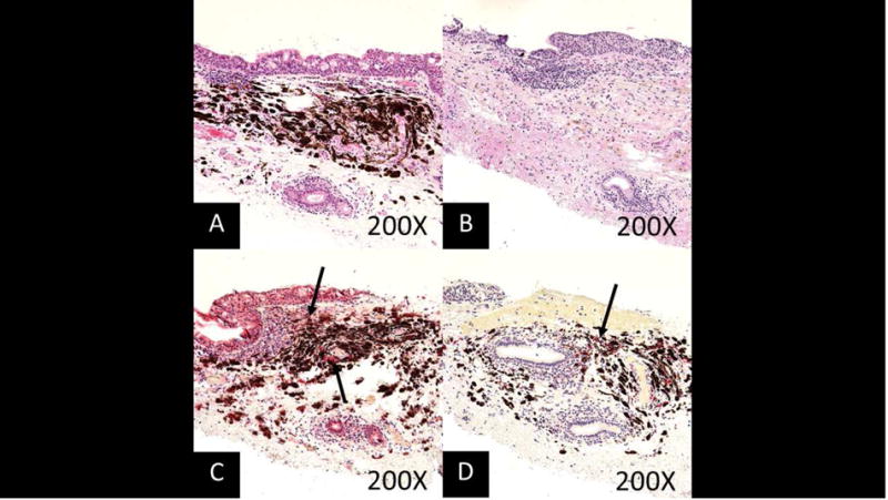
Methods: Chart review of patients identified from a database search of the Florida Lions Ocular Pathology Laboratory records. Pathologic diagnoses were confirmed by 2 pathologists (S.R.D. and G.E.). All specimens were bleached and, tissue permitting, stained using SOX10 (MilliporeSigma, Darmstadt, Germany) and CD68 (Leica Biosystems, Nussloch, Germany).
Main outcome measures: Clinical characteristics, pathologic features, and clinical course.
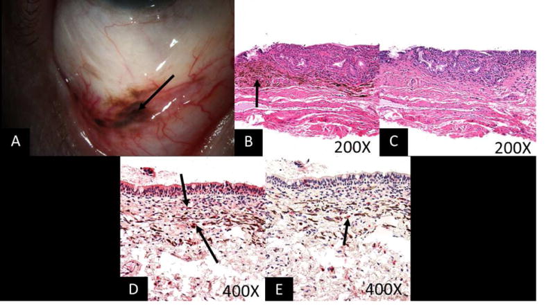
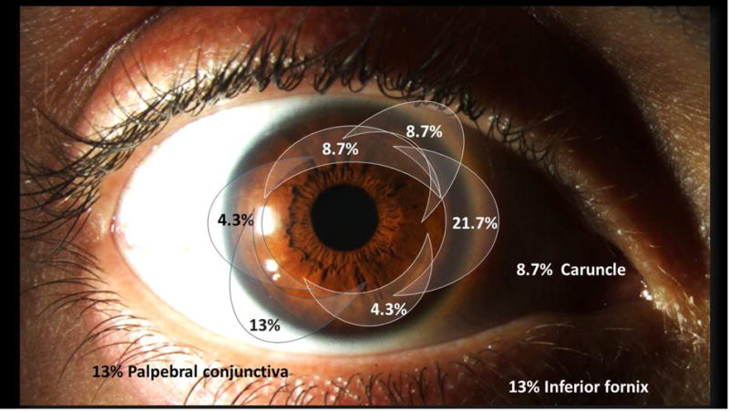
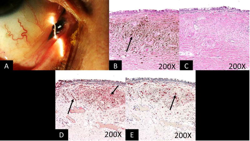
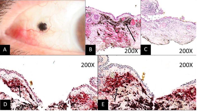
Results: Mean age of the population was 55±15 years; 71.4% (n = 15) were white and 57.1% (n = 12) were men. One patient had 3 lesions, for a total of 23 lesions examined. Clinically, 13 lesions were on the bulbar conjunctiva, 3 were on the tarsal conjunctiva, 3 were in the fornix, 2 were caruncular, 1 was episcleral, and 1 was at the limbus. Before excision, 8 patients were thought to have primary acquired melanosis, 4 with concern for primary conjunctival melanoma, and 1 thought to have metastatic disease from a plantar melanoma. Five lesions were thought to be benign, and in 8 patients, the lesions were identified incidentally after other ocular surgeries, with no diagnosis of the lesions before excision. Pathologic features were consistent with simple blue nevi in 21 lesions and cellular blue nevus in 2 lesions. No malignant transformations were noted in any patient over the mean 20.2-month follow-up period (range, 2 weeks-103 months).
Conclusions: Blue nevus is a rare deeply pigmented congenital melanocytic lesion with a benign clinical course that can appear clinically similar to primary acquired melanosis or melanoma.
Copyright © 2018 American Academy of Ophthalmology. All rights reserved.
Conflict of interest statement
Figures
Similar articles
-
[Blue nevi of the conjunctiva].Klin Monbl Augenheilkd. 1994 Oct;205(4):242-3. doi: 10.1055/s-2008-1045521. Klin Monbl Augenheilkd. 1994. PMID: 7823524 German.
-
Usefulness of a red chromagen in the diagnosis of melanocytic lesions of the conjunctiva.JAMA Ophthalmol. 2014 May;132(5):622-9. doi: 10.1001/jamaophthalmol.2013.8216. JAMA Ophthalmol. 2014. PMID: 24626521
-
Blue nevi of the palpebral conjunctiva: report of 2 cases and review of literature.Orbit. 2022 Oct;41(5):527-534. doi: 10.1080/01676830.2022.2065315. Epub 2022 Apr 28. Orbit. 2022. PMID: 35482915 Review.
-
Benign conjunctival melanocytic lesions. Clinicopathologic features.Ophthalmology. 1989 Apr;96(4):436-61. doi: 10.1016/s0161-6420(89)32878-8. Ophthalmology. 1989. PMID: 2657539 Review.
-
Incidence of melanocytic lesions of the conjunctiva in a review of 10 675 ophthalmic specimens.Int J Surg Pathol. 2010 Feb;18(1):60-3. doi: 10.1177/1066896908319775. Epub 2008 Jul 8. Int J Surg Pathol. 2010. PMID: 18611943
Cited by
-
Melanocytoma of the eyelid: Case report and introduction of new nomenclature.Am J Ophthalmol Case Rep. 2024 Apr 16;34:102059. doi: 10.1016/j.ajoc.2024.102059. eCollection 2024 Jun. Am J Ophthalmol Case Rep. 2024. PMID: 38690089 Free PMC article.
-
Conjunctival Nevus.Curr Ophthalmol Rep. 2023 Dec;11(4):104-112. doi: 10.1007/s40135-023-00315-w. Epub 2023 Jul 19. Curr Ophthalmol Rep. 2023. PMID: 38390435 Free PMC article.
-
The use of high resolution optical coherence tomography (HR-OCT) in the diagnosis of ocular surface masqueraders.Ocul Surf. 2022 Apr;24:74-82. doi: 10.1016/j.jtos.2022.02.003. Epub 2022 Feb 26. Ocul Surf. 2022. PMID: 35231640 Free PMC article. Review.
-
Recurrent melanoma arising from sclera.Am J Ophthalmol Case Rep. 2022 May 5;27:101562. doi: 10.1016/j.ajoc.2022.101562. eCollection 2022 Sep. Am J Ophthalmol Case Rep. 2022. PMID: 35677815 Free PMC article.
-
A Case of Nevus of Ota in Trinidad and Tobago.Cureus. 2025 Jan 31;17(1):e78311. doi: 10.7759/cureus.78311. eCollection 2025 Jan. Cureus. 2025. PMID: 39897319 Free PMC article.
References
-
- Kirzhner M, Jakobiec FA, Kim N. Focal Blue Nevus of the Eyelid Margin (Mucocutaneous Junction): A Report of a Unique Case With a Review of the Literature. Ophthalmic Plastic & Reconstructive Surgery. 2011;27(5):338–42. - PubMed
-
- Murali R, McCarthy SW, Scolyer RA. Blue nevi and related lesions: a review highlighting atypical and newly described variants, distinguishing features and diagnostic pitfalls. Adv Anat Pathol. 2009;16(6):365–82. - PubMed
-
- Zembowicz A, Phadke PA. Blue nevi and variants: an update. Archives of pathology & laboratory medicine. 2011;135(3):327–36. - PubMed
-
- Rodriguez HA, Ackerman LV. Cellular blue nevus. Clinicopathologic study of forty-five cases. Cancer. 1968;21(3):393–405. - PubMed
-
- Jakobiec FA, Colby K, Bajart AM, et al. Immunohistochemical studies of atypical conjunctival melanocytic nevi. Archives of ophthalmology. 2009;127(8):970–80. - PubMed
Publication types
MeSH terms
Grants and funding
LinkOut - more resources
Full Text Sources
Other Literature Sources
Research Materials
