Proliferative verrucous leukoplakia misdiagnosed as oral leukoplakia
- PMID: 29551871
- PMCID: PMC5846249
- DOI: 10.4103/jisp.jisp_189_17
Proliferative verrucous leukoplakia misdiagnosed as oral leukoplakia
Abstract
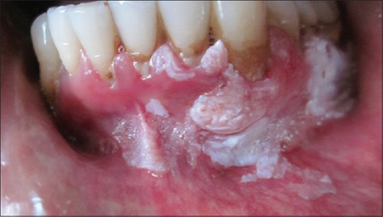
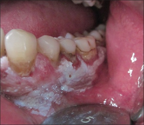
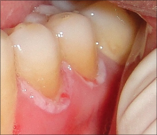
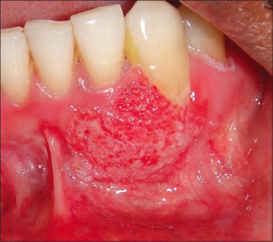
Correct diagnosis of white lesions of the oral cavity is sometimes difficult, because some oral white lesions behave differently and tend to change their appearance with time. Clinicians often wrongly diagnose such lesions as oral leukoplakias and treat simply. Lesions recur and turn malignant. Proliferative verrucous leukoplakia (PVL) is a distinct form of oral leukoplakia characterized by a high recurrence rate and high rate of transformation into oral squamous cell carcinoma. We present herein a case of PVL which was misdiagnosed as oral leukoplakia and progressed to oral carcinoma.
Keywords: Malignant; multifactorial; multifocal; proliferative verrucous leukoplakia; squamous cell carcinoma.
Conflict of interest statement
There are no conflicts of interest.
Figures
Similar articles
-
Proliferative verrucous leukoplakia: an elusive disorder.J Evid Based Dent Pract. 2014 Jun;14 Suppl:147-53.e1. doi: 10.1016/j.jebdp.2014.04.005. Epub 2014 Apr 5. J Evid Based Dent Pract. 2014. PMID: 24929599 Review.
-
Proliferative verrucous leukoplakia: Case study of 24 years and outcome of treatment with CO2 laser.Clin Adv Periodontics. 2024 Oct 24. doi: 10.1002/cap.10320. Online ahead of print. Clin Adv Periodontics. 2024. PMID: 39447057
-
Proliferative verrucous leukoplakia and its related lesions.Oral Oncol. 1999 Jul;35(4):354-9. doi: 10.1016/s1368-8375(99)00007-x. Oral Oncol. 1999. PMID: 10645398 Review.
-
Oral Carcinoma Arising Under Implant-Supported Prosthesis: Progression of Proliferative Verrucous Leukoplakia Initially Mimicking Lichen Planus.J Oral Implantol. 2024 Aug 1;50(4):397-400. doi: 10.1563/aaid-joi-D-24-00056. J Oral Implantol. 2024. PMID: 38742460
-
[Proliferative verrucous leukoplakia. Report of five cases].Mund Kiefer Gesichtschir. 2003 May;7(3):164-70. doi: 10.1007/s10006-003-0472-1. Epub 2003 May 1. Mund Kiefer Gesichtschir. 2003. PMID: 12764683 German.
Cited by
-
Development and Evaluation of an Automated Multimodal Mobile Detection of Oral Cancer Imaging System to Aid in Risk-Based Management of Oral Mucosal Lesions.Cancer Prev Res (Phila). 2025 Apr 1;18(4):197-207. doi: 10.1158/1940-6207.CAPR-24-0253. Cancer Prev Res (Phila). 2025. PMID: 39817650 Free PMC article.
-
Oral Potentially Malignant Disorders: Etiology, Pathogenesis, and Transformation Into Oral Cancer.Front Pharmacol. 2022 Apr 20;13:825266. doi: 10.3389/fphar.2022.825266. eCollection 2022. Front Pharmacol. 2022. PMID: 35517828 Free PMC article. Review.
-
Proliferative verrucous leukoplakia, an enigma to the pathologists: Report of two cases.SAGE Open Med Case Rep. 2024 Mar 11;12:2050313X241236335. doi: 10.1177/2050313X241236335. eCollection 2024. SAGE Open Med Case Rep. 2024. PMID: 38476568 Free PMC article.
-
Gingival Proliferative Verrucous Leukoplakia and Cancer: A Systematic Review With Meta-Analysis.Oral Dis. 2025 Jan;31(1):50-58. doi: 10.1111/odi.15142. Epub 2024 Oct 9. Oral Dis. 2025. PMID: 39385511 Free PMC article.
References
-
- Warnakulasuriya S, Johnson NW, van der Waal I. Nomenclature and classification of potentially malignant disorders of the oral mucosa. J Oral Pathol Med. 2007;36:575–80. - PubMed
-
- Hansen LS, Olson JA, Silverman S., Jr Proliferative verrucous leukoplakia. A long-term study of thirty patients. Oral Surg Oral Med Oral Pathol. 1985;60:285–98. - PubMed
-
- Salati NA. Clinic-pathologic evaluation and medical treatment of oral leukoplakia. Int J Pharm Sci Invent. 2014;3:7–12.
-
- Seoane J, Varela-Centelles PI, Walsh TF, Lopez-Cedrun JL, Vazquez I. Gingival squamous cell carcinoma: Diagnostic delay or rapid invasion? J Periodontol. 2006;77:1229–33. - PubMed
-
- Gandolfo S, Castellani R, Pentenero M. Proliferative verrucous leukoplakia: A potentially malignant disorder involving periodontal sites. J Periodontol. 2009;80:274–81. - PubMed