Review
. 2018 Mar 1:12:1179069518759332.
doi: 10.1177/1179069518759332. eCollection 2018.

Untangling Cortical Complexity During Development

Affiliations
Review

Untangling Cortical Complexity During Development

Tanzila Mukhtar et al. J Exp Neurosci. .

Abstract

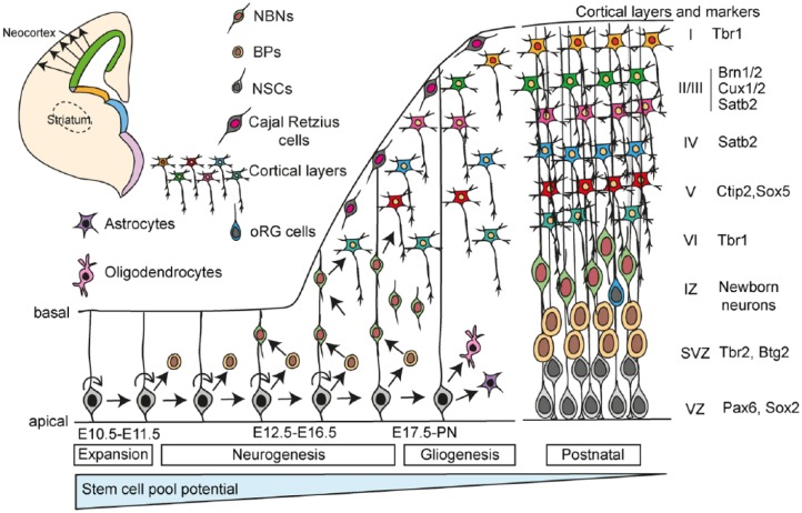
The cerebral cortex is composed of billions of morphologically and functionally distinct neurons. These neurons are produced and organized in a regimental fashion during development. The ability of neurons to encode and elicit complex cognitive and motor functions depends on their precise molecular processes, identity, and connectivity established during development. Elucidating the cellular and molecular mechanisms that regulate development of the neocortex has been a challenge for many years. The cerebral cortical neuronal subtypes are classified based on morphology, function, intrinsic synaptic properties, location, connectivity, and marker gene expression. Development of the neocortex requires an orchestration of a series of processes including the appropriate determination, migration and positioning of the neurons, acquisition of layer-specific transcriptional hallmarks, and formation of precise axonal projections and networks. Historically, fate mapping, genome-wide analysis, and transcriptome profiling have provided many opportunities for the characterization of neuronal subtypes. During the course of this review, we will address the regimental organization of the cerebral cortex, dissect the cellular subtypes that contribute to cortical complexity, and outline their molecular hallmarks to understand cellular diversity in the cerebral cortex with a focus on the excitatory neurons.

Keywords: Brain development; cerebral cortex; neural stem cells; neurogenesis.


Conflict of interest statement

Declaration of conflicting interests:The author(s) declared no potential conflicts of interest with respect to the research, authorship, and/or publication of this article.

Figures

Figure 1.
Figure 1.
Types of NSC divisions in the ventricular zone are determined by spindle orientation and the inheritance of cell fate determinants. Symmetric divisions generate 2 NSCs, whereas asymmetric division generates 1 NSC and 1 differentiating daughter cell. During neural expansion, most divisions are symmetric, whereas during neurogenesis, most divisions are asymmetric. BP indicates basal progenitor; NSC, neural stem cell.
Figure 2.
Figure 2.
Scheme illustrating the composition and laminar organization of the developing human cortex, in comparison with mouse cortex. The human cerebral cortex develops in a similar fashion to that of the mouse. One exception is the expansion of the subventricular zone (SVZ) to form the outer SVZ (oSVZ). The oSVZ in humans is the main zone of amplification. In addition to the neural stem cells (NSCs) and basal progenitors (BPs) of the developing mouse cerebral cortex, the human has addition progenitors, outer radial glial cells. CP indicates cortical plate; IZ, intermediate zone; MZ, marginal zone; SP, subplate; SVZ, subventricular zone; VZ, ventricular zone.
Figure 3.
Figure 3.
Different models of neuronal subtype specification in developing neocortex. (A) In the common progenitor model, a single type of multipotent NSC sequentially gives rise to all neuronal subtypes during the course of development. Overtime, the fate potential of this NSC becomes increasingly restricted. Fate of the neuron is specified based on its birth date. (B) In the multiple progenitor model, multiple types of NSCs coexist and are, to some degree, predetermined to give rise to specific and restricted neuronal subtypes. Fate of the neuron is specified by the NSC type in this model. BP indicates basal progenitor; NSC, neural stem cell.
Figure 4.
Figure 4.
Origin of excitatory projection neurons of the cerebral cortex. Excitatory projection neurons originate from the ventricular zone of the dorsal telencephalon and migrate radially to the cortical plate. Inhibitory interneurons originate from the ventral telencephalon, especially from the MGE, AEP/POA., AEP/POA indicates anterior entopeduncular area of the subpallium/preoptic area; LGE, lateral ganglionic eminence; LP, lateral pallium; MGE, medial ganglionic eminence.
Figure 5.
Figure 5.
Systematic formation of isocortex layers in the dorsal telencephalon. During early stages of cerebral cortical development (embryonic days E10.5-E11.5), NSCs predominantly undergo symmetric cells divisions to expand the NSC pool. This phase is referred to as the expansion phase. The first neurons to be formed are generated by direct neurogenesis of the NSCs. The Cajal-Retzius cells populate layer I of the isocortex and play important roles in establishing cortical architecture. During late embryogenesis (E12-E16.5), NSCs undergo increasingly more asymmetric divisions to generate 1 NSC (self-renewal) and 1 BP. The BPs generate the neurons. This is the neurogenic phase. Neurons are generated in a sequential, inside-out fashion and are specified by different transcription factors, some of which are shown. At later stages of development, NSCs generate the other cell types of the brain including astrocytes, oligodendrocytes, and ependymal cells (not shown). This is referred to as the gliogenic phase. The potential of the NSC pool reduces over time during development. This does not exclude that multiple restricted stem cells become activated and are lost at different times during cortical development. BPs indicate basal progenitors; IZ, intermediate zone; NBNs, newborn neurons; NSCs, neural stem cells; SVZ, subventricular zone; VZ, ventricular zone.

References

    1. Tam PP, Loebel DA. Gene function in mouse embryogenesis: get set for gastrulation. Nat Rev Genet. 2007;8:368–381. - PubMed
    1. Lee HK, Lee HS, Moody SA. Neural transcription factors: from embryos to neural stem cells. Mol Cells. 2014;37:705–712. - PMC - PubMed
    1. Paridaen JT, Huttner WB. Neurogenesis during development of the vertebrate central nervous system. EMBO Rep. 2014;15:351–364. - PMC - PubMed
    1. Zhadanov AB, Provance DW, Jr, Speer CA, et al. Absence of the tight junctional protein AF-6 disrupts epithelial cell-cell junctions and cell polarity during mouse development. Curr Biol. 1999;9:880–888. - PubMed
    1. Gal JS, Morozov YM, Ayoub AE, Chatterjee M, Rakic P, Haydar TF. Molecular and morphological heterogeneity of neural precursors in the mouse neocortical proliferative zones. J Neurosci. 2006;26:1045–1056. - PMC - PubMed

LinkOut - more resources