

Making Thymus Visible: Understanding T-Cell Development from a New Perspective
- PMID: 29552011
- PMCID: PMC5840141
- DOI: 10.3389/fimmu.2018.00375
Making Thymus Visible: Understanding T-Cell Development from a New Perspective
Abstract
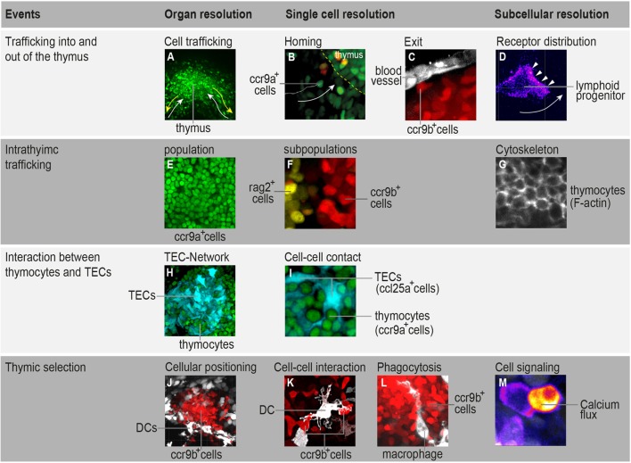
T-cell development is coupled with a highly ordered migratory pattern. Lymphoid progenitors must follow a precise journey; starting from the hematopoietic tissue, they move toward the thymus and then migrate into and out of distinct thymic microenvironments, where they receive signals and cues required for their differentiation into naïve T-cells. Knowing where, when, and how these cells make directional "decisions" is key to understanding T-cell development. Such insights can be gained by directly observing developing T-cells within their environment under various conditions and following specific experimental manipulations. In the last decade, several model systems have been developed to address temporal and spatial aspects of T-cell development using imaging approaches. In this perspective article, we discuss the advantages and limitations of these systems and highlight a particularly powerful in vivo model that has been recently established. This model system enables the migratory behavior of all thymocytes to be studied simultaneously in a noninvasive and quantitative manner, making it possible to perform systems-level studies that reveal fundamental principles governing T-cell dynamics during development and in disease.
Keywords: chemokines; imaging; medaka; thymus; zebrafish.
Figures
Similar articles
-
Noninvasive In Toto Imaging of the Thymus Reveals Heterogeneous Migratory Behavior of Developing T Cells.J Immunol. 2015 Sep 1;195(5):2177-86. doi: 10.4049/jimmunol.1500361. Epub 2015 Jul 17. J Immunol. 2015. PMID: 26188059
-
Molecular mechanisms governing thymocyte migration: combined role of chemokines and extracellular matrix.J Leukoc Biol. 2004 Jun;75(6):951-61. doi: 10.1189/jlb.1003455. Epub 2004 Mar 12. J Leukoc Biol. 2004. PMID: 15020651 Review.
-
RasGRP1 and RasGRP3 Are Required for Efficient Generation of Early Thymic Progenitors.J Immunol. 2016 Sep 1;197(5):1743-53. doi: 10.4049/jimmunol.1502107. Epub 2016 Jul 27. J Immunol. 2016. PMID: 27465532
-
T-cell selection in the thymus: a spatial and temporal perspective.Immunol Rev. 2016 May;271(1):114-26. doi: 10.1111/imr.12398. Immunol Rev. 2016. PMID: 27088910 Free PMC article. Review.
-
Notch1 deficiency alters the migratory behavior of developing T cells and calcium signaling in the thymus of medaka.Eur J Immunol. 2022 Feb;52(2):261-269. doi: 10.1002/eji.202149512. Epub 2021 Nov 17. Eur J Immunol. 2022. PMID: 34731490
Cited by
-
Whole-transcriptome profiling reveals potential biomarkers for the reversal of thymic epithelial cell senescence by umbilical cord mesenchymal stem cells.Aging (Albany NY). 2024 Apr 17;16(8):7009-7021. doi: 10.18632/aging.205738. Epub 2024 Apr 17. Aging (Albany NY). 2024. PMID: 38637117 Free PMC article.
-
Nano-Sampling and Reporter Tools to Study Metabolic Regulation in Zebrafish.Front Cell Dev Biol. 2019 Feb 19;7:15. doi: 10.3389/fcell.2019.00015. eCollection 2019. Front Cell Dev Biol. 2019. PMID: 30873407 Free PMC article.
-
Nck1 regulates the in vitro development of human regulatory T cells through AKT pathway.Clin Exp Immunol. 2025 Jan 21;219(1):uxaf011. doi: 10.1093/cei/uxaf011. Clin Exp Immunol. 2025. PMID: 39963999
-
Intravital two-photon microscopy of the native mouse thymus.PLoS One. 2024 Aug 1;19(8):e0307962. doi: 10.1371/journal.pone.0307962. eCollection 2024. PLoS One. 2024. PMID: 39088574 Free PMC article.
-
Inflammatory Responses during Tumour Initiation: From Zebrafish Transgenic Models of Cancer to Evidence from Mouse and Man.Cells. 2020 Apr 20;9(4):1018. doi: 10.3390/cells9041018. Cells. 2020. PMID: 32325966 Free PMC article. Review.
References
Publication types
MeSH terms
LinkOut - more resources
Full Text Sources
Other Literature Sources