

Gastric cancer patients have elevated plasmacytoid and CD1c+ dendritic cells in the peripheral blood
- PMID: 29552142
- PMCID: PMC5840537
- DOI: 10.3892/ol.2018.7990
Gastric cancer patients have elevated plasmacytoid and CD1c+ dendritic cells in the peripheral blood
Abstract
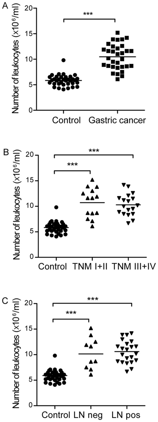
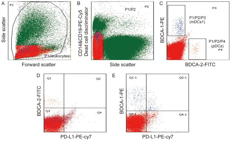
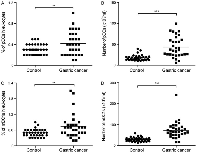
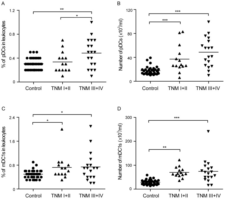
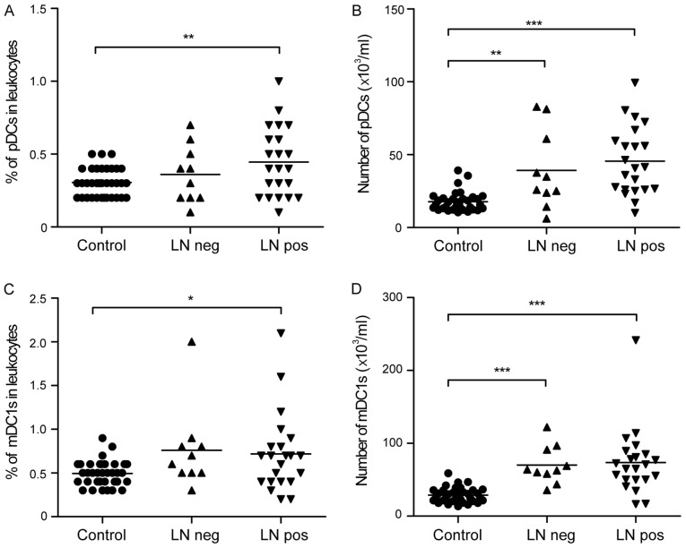
Dendritic cells (DCs) are important in tumor immunology. Identifying DC subset markers in the peripheral blood, which are informative for gastric cancer stages, is not only useful for prognosis but may also provide mechanistic insights into processes facilitating therapy. The present study investigated plasmacytoid dendritic cells (pDCs) and myeloid CD1c+ dendritic cells (mDC1s) in the peripheral blood of patients with gastric cancer and healthy controls using flow cytometry. Using peripheral DC staining and subset analysis, patients with gastric cancer were identified to have substantially higher numbers of peripheral pDCs and mDC1s. In addition, there was a trend of elevated circulating pDCs with advanced stages and lymph node metastasis in gastric cancer, whereas no differences in circulating mDC1s were observed among the various groups. The results suggested that circulating pDCs are a positive prognostic indicator in patients with gastric cancer of different stages and highlighted the critical role of pDCs immunity in the development of gastric cancer.
Keywords: dendritic cell; gastric cancer; myeloid dendritic cell; plasmacytoid dendritic cell.
Figures
Similar articles
-
Phenotypic and Transcriptomic Analysis of Peripheral Blood Plasmacytoid and Conventional Dendritic Cells in Early Drug Naïve Rheumatoid Arthritis.Front Immunol. 2018 May 9;9:755. doi: 10.3389/fimmu.2018.00755. eCollection 2018. Front Immunol. 2018. PMID: 29867920 Free PMC article.
-
Prognostic impact of high levels of circulating plasmacytoid dendritic cells in breast cancer.J Transl Med. 2016 May 28;14(1):151. doi: 10.1186/s12967-016-0905-x. J Transl Med. 2016. PMID: 27234566 Free PMC article.
-
Acute aerobic exercise induces a preferential mobilisation of plasmacytoid dendritic cells into the peripheral blood in man.Physiol Behav. 2018 Oct 1;194:191-198. doi: 10.1016/j.physbeh.2018.05.012. Epub 2018 May 31. Physiol Behav. 2018. PMID: 29763678
-
Primary Human Blood Dendritic Cells for Cancer Immunotherapy-Tailoring the Immune Response by Dendritic Cell Maturation.Biomedicines. 2015 Dec 2;3(4):282-303. doi: 10.3390/biomedicines3040282. Biomedicines. 2015. PMID: 28536413 Free PMC article. Review.
-
Tumor-forming plasmacytoid dendritic cells in acute myelocytic leukemia: a report of three cases and literature review.Int J Clin Exp Pathol. 2017 Jul 1;10(7):7285-7291. eCollection 2017. Int J Clin Exp Pathol. 2017. PMID: 31966568 Free PMC article. Review.
Cited by
-
The Multifaceted Functionality of Plasmacytoid Dendritic Cells in Gastrointestinal Cancers: A Potential Therapeutic Target?Cancers (Basel). 2024 Jun 13;16(12):2216. doi: 10.3390/cancers16122216. Cancers (Basel). 2024. PMID: 38927922 Free PMC article. Review.
-
The Interplay between Circulating Tumor Cells and the Immune System: From Immune Escape to Cancer Immunotherapy.Diagnostics (Basel). 2018 Aug 30;8(3):59. doi: 10.3390/diagnostics8030059. Diagnostics (Basel). 2018. PMID: 30200242 Free PMC article. Review.
-
Advances in Human Dendritic Cell-Based Immunotherapy Against Gastrointestinal Cancer.Front Immunol. 2022 May 10;13:887189. doi: 10.3389/fimmu.2022.887189. eCollection 2022. Front Immunol. 2022. PMID: 35619702 Free PMC article. Review.
-
CD1C is associated with breast cancer prognosis and immune infiltrates.BMC Cancer. 2023 Feb 8;23(1):129. doi: 10.1186/s12885-023-10558-2. BMC Cancer. 2023. PMID: 36755259 Free PMC article.
-
Upregulation of ZHX2 predicts poor prognosis and is correlated with immune infiltration in gastric cancer.FEBS Open Bio. 2021 Jun;11(6):1785-1798. doi: 10.1002/2211-5463.13160. Epub 2021 May 24. FEBS Open Bio. 2021. PMID: 33837660 Free PMC article.
References
-
- Kashimura S, Saze Z, Terashima M, Soeta N, Ohtani S, Osuka F, Kogure M, Gotoh M. CD83(+) dendritic cells and Foxp3(+) regulatory T cells in primary lesions and regional lymph nodes are inversely correlated with prognosis of gastric cancer. Gastric Cancer. 2012;15:144–153. doi: 10.1007/s10120-011-0090-9. - DOI - PubMed
-
- Tsukayama S, Omura K, Yoshida K, Tanaka Y, Watanabe G. Prognostic value of CD83-positive mature dendritic cells and their relation to vascular endothelial growth factor in advanced human gastric cancer. Oncol Rep. 2005;14:369–375. - PubMed
LinkOut - more resources
Full Text Sources
Other Literature Sources