. 2018 Apr;15(4):5611-5619.
doi: 10.3892/ol.2018.8050. Epub 2018 Feb 14.

MicroRNA-10a/b are regulators of myeloid differentiation and acute myeloid leukemia

Affiliations

MicroRNA-10a/b are regulators of myeloid differentiation and acute myeloid leukemia

Laixi Bi et al. Oncol Lett. 2018 Apr.

Abstract

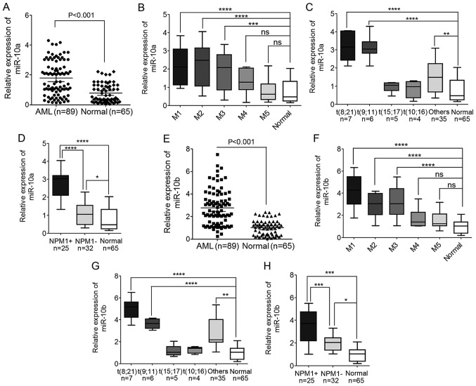
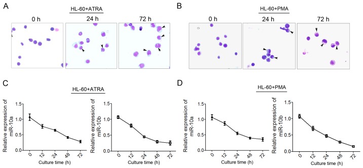
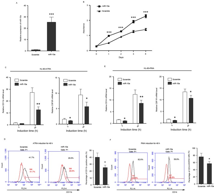
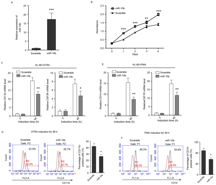
MicroRNAs (miRs) have been demonstrated to perform important roles in normal hematopoiesis and leukemogenesis. Accumulating evidence suggests that miR-10a and miR-10b may behave as novel oncogenes or tumor suppressors in human cancer. The present study reported the function of the miR-10 family in myeloid differentiation and acute myeloid leukemia (AML). The levels of miR-10a/b expression were increased in AML cases compared with normal controls, particularly in M1, M2 and M3 subtypes. The levels of miR-10a/b expression were also upregulated in patients with nucleophosmin-mutated AML and AML patients with t(8;21) and t(9;11), compared with the normal control. In addition, the role of miR-10a/b in regulating myeloid differentiation and leukemogenesis was investigated. The results indicated that miR-10a/b expression was able to promote the proliferation of human promyelocytic leukemia cells, while suppressing the granulocytic and monocytic differentiation of the leukemia cells. These findings suggested that abnormal high expression of miR-10a/b may result in unlimited proliferation of immature blood progenitors and repression of mature blood cell differentiation and maturation, thus leading to the occurrence of AML. miR-10a/b may be developed as novel therapeutic targets for the treatment of AML.

Keywords: acute myeloid leukemia; microRNA-10a/b; myeloid differentiation.


Figures

Figure 1.
Figure 1.
Levels of miR-10a and miR-10b expression are significantly increased in AML cases. (A) The expression level of miR-10a in PBMNCs derived from 89 patients with AML and 65 healthy donors detected by TaqMan quantitative polymerase chain reaction. (B) The expression level of miR-10a in different AML subtypes, from M1 to M5. (C) The expression level of miR-10a in different WHO subtypes of AML patients compared with the normal controls. (D) The expression level of miR-10a in AML patients with mutated and wild-type NPM1 compared with the normal controls. (E) The expression level of miR-10b in PBMNCs derived from 89 AML patients and 65 healthy donors. (F) The expression level of miR-10b in different AML subtypes, from M1 to M5. (G) The expression level of miR-10b in different WHO subtypes of AML patients compared with the normal controls. (H) The expression level of miR-10b in AML patients with mutated and wild-type NPM1 compared with the normal controls. *P<0.05, **P<0.01, ***P<0.001, ****P<0.0001; ns, not significant. AML, acute myeloid leukemia; miR, microRNA; NPM1, nucleophosmin; PBMNC, peripheral blood mononuclear cell; WHO, World Health Organization; NPM1+, NPM1 mutated; NPM1-, wild-type NPM1.
Figure 2.
Figure 2.
Decreased miR-10a/b expression during granulocytic and monocytic differentiation of HL-60 cells. (A and B) May-Grünwald Giemsa staining of HL-60 cells that were induced by ATRA or PMA for 0, 24 and 72 h, respectively. The differentiated macrophages and monocytes were indicated with arrows. (C) Changes in expression level of miR-10a and miR-10b during ATRA-induced HL-60 granulocytic differentiation at 12, 24, 48 and 72 h post-induction. (D) Changes in the level of (C) miR-10a and (D) miR-10b expression during PMA-induced monocytic differentiation. Data are presented as the mean ± standard deviation. miR, microRNA; ATRA, all-trans-retinoic acid; PMA, phorbol myristate acetate.
Figure 3.
Figure 3.
miR-10a promotes the proliferation of HL-60 cells, while inhibits the differentiation of HL-60 cells to granulocytes and monocytes. (A) miR-10a is overexpressed in HL-60 cells as confirmed by quantitative polymerase chain reaction. (B) The growth of HL-60 cells at day 0, 1, 2, 3 and 4 post-transfection, which was detected by Cell Counting Kit-8 assay. (C) The expression level of granulocytic marker CD11b and CSF3R in untreated and ATRA-treated (duration, 48 h) cells that overexpress miR-10a. (D) CD11b FACS analysis showed that overexpression of miR-10a was able to delay ATRA-induced granulocytic differentiation of HL-60 cells. The percentage of CD11b-positive cells was calculated using the black traces referring to cells stained with CD11b antibody against the red traces referring to the same cells not stained with CD11b antibody. (E) The relative expression of monocytic marker CD14 and CSF1R in untreated and PMA-treated (duration, 48 h) cells that overexpress miR-10a. (F) CD14 FACS analysis showed that overexpression of miR-10a delayed PMA-induced monocytic differentiation of HL-60 cells. The percentage of CD14-positive cells was calculated in the same way. Data are presented as the mean ± standard deviation (n=3). All the comparisons are made between scramble and miR-10a groups. *P<0.05; **P<0.01; ***P<0.001. miR, microRNA; CSF3R, colony-stimulating factor 3 receptor; ATRA, all-trans-retinoic acid; PMA, phorbol myristate acetate; CD, cluster of differentiation; CSF1R, colony-stimulating factor 1 receptor; FACS, fluorescence-activated cell sorting.
Figure 4.
Figure 4.
miR-10b promotes the proliferation of HL-60 cells, while inhibits the differentiation of HL-60 cells to granulocytes and monocytes. (A) The overexpression of miR-10b in HL-60 cells as confirmed by quantitative polymerase chain reaction. (B) The growth of HL-60 cells at day 0, 1, 2, 3 and 4 post-transfection as detected by Cell Counting Kit-8 assay. (C) The expression level of granulocytic marker CD11b and CSF3R in untreated and ATRA-treated (duration, 48 h) cells that overexpress miR-10b. (D) CD11b FACS analysis showed that enforced expression of miR-10b delayed ATRA-induced granulocytic differentiation of HL-60 cells. (E) The relative expression of monocytic marker CD14 and CSF1R in untreated and PMA-treated (duration, 48 h) cells that overexpress miR-10b. (F) CD14 FACS analysis showed that enforced expression of miR-10b delayed PMA-induced monocytic differentiation of HL-60 cells. Data are presented as the mean ± standard deviation (n=3); *P<0.05; **P<0.01; ***P<0.001. All the comparisons are made between scramble and miR-10a groups. miR, microRNA; CSF3R, colony-stimulating factor 3 receptor; CD, cluster of differentiation; CSF1R, colony-stimulating factor 1 receptor; ATRA, al-trans-retinoic acid; PMA, phorbol myristate acetate; FACS, fluorescence-activated cell sorting.

References

    1. Birbrair A, Frenette PS. Niche heterogeneity in the bone marrow. Ann N Y Acad Sci. 2016;1370:82–96. doi: 10.1111/nyas.13016. - DOI - PMC - PubMed
    1. Morrison SJ, Kimble J. Asymmetric and symmetric stem-cell divisions in development and cancer. Nature. 2006;441:1068–1074. doi: 10.1038/nature04956. - DOI - PubMed
    1. Falini B, Tiacci E, Martelli MP, Ascani S, Pileri SA. New classification of acute myeloid leukemia and precursor-related neoplasms: Changes and unsolved issues. Discov Med. 2010;10:281–292. - PubMed
    1. Jemal A, Thomas A, Murray T, Thun M. Cancer statistics, 2002. CA Cancer J Clin. 2002;52:23–47. doi: 10.3322/canjclin.52.1.23. - DOI - PubMed
    1. Döhner H, Weisdorf DJ, Bloomfield CD. Acute myeloid leukemia. N Engl J Med. 2015;373:1136–1152. doi: 10.1056/NEJMra1406184. - DOI - PubMed