

Anti-oncogenic activity of Chibby in the development of human nasopharyngeal carcinoma
- PMID: 29552214
- PMCID: PMC5840504
- DOI: 10.3892/ol.2018.8009
Anti-oncogenic activity of Chibby in the development of human nasopharyngeal carcinoma
Abstract
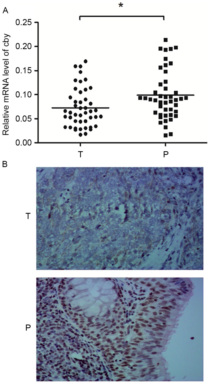
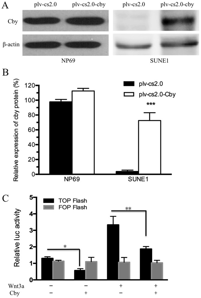
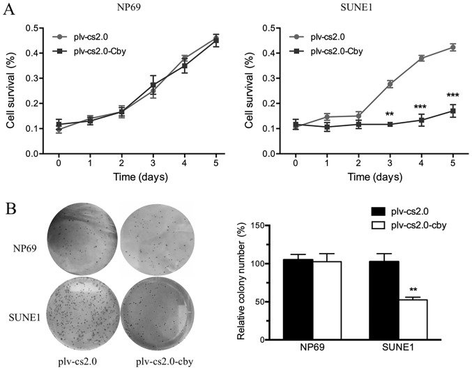
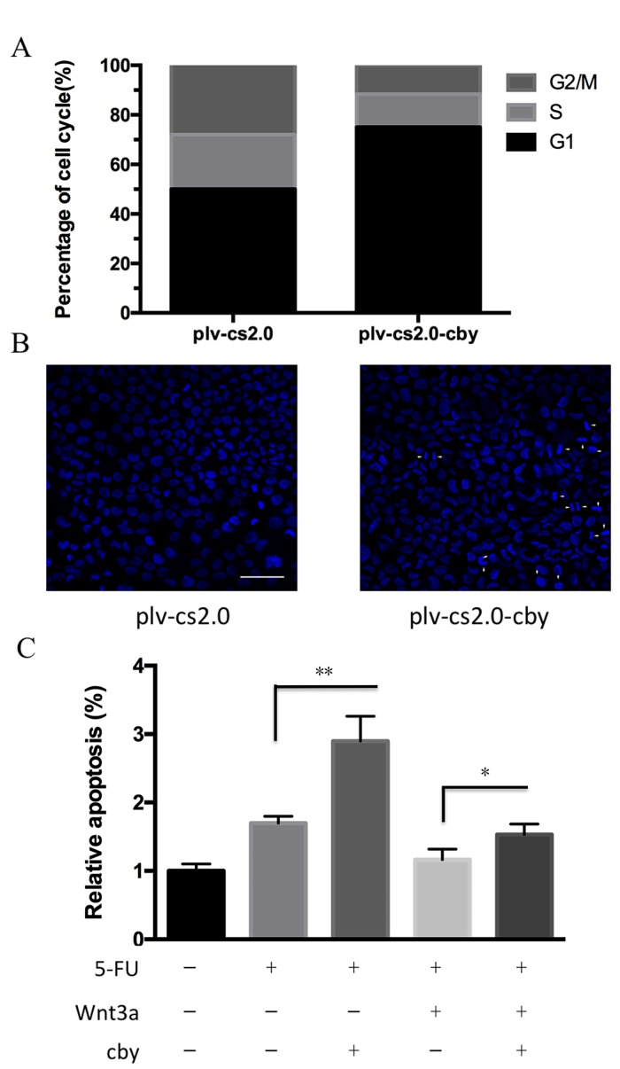
The Wnt/β-catenin pathway serves important roles in cancer development. The expression and function of Chibby (Cby), as a direct antagonist of β-catenin, in nasopharyngeal carcinoma (NPC) has not been fully investigated. The present study revealed that the mRNA and protein expression of Cby was significantly lower in NPC tissue than in the adjacent normal tissue. Low expression of Cby was significantly associated with the tumor and the clinical staging. Furthermore, Cby overexpression inhibited the proliferation of human NPC SUNE1 cells and induced cell cycle arrest. In addition, Cby overexpression also significantly enhanced the susceptibility of SUNE1 cells to apoptosis. These results indicated that Cby might serve as an anti-oncogenic gene in the development of NPC and could represent a potential therapeutic target for the human NPC therapy.
Keywords: Chibby; Wnt/β-catenin; apoptosis; cell cycle; nasopharyngeal carcinoma.
Figures
Similar articles
-
Chibby suppresses aerobic glycolysis and proliferation of nasopharyngeal carcinoma via the Wnt/β-catenin-Lin28/let7-PDK1 cascade.J Exp Clin Cancer Res. 2018 May 15;37(1):104. doi: 10.1186/s13046-018-0769-4. J Exp Clin Cancer Res. 2018. PMID: 29764469 Free PMC article.
-
Downregulated Chibby in laryngeal squamous cell carcinoma with increased expression in laryngeal carcinoma Hep-2 cells.Oncol Rep. 2014 Nov;32(5):1947-56. doi: 10.3892/or.2014.3451. Epub 2014 Aug 29. Oncol Rep. 2014. PMID: 25175341
-
Chibby is a weak regulator of β-catenin activity in gastric epithelium.J Cell Physiol. 2019 Feb;234(2):1871-1879. doi: 10.1002/jcp.27062. Epub 2018 Jul 31. J Cell Physiol. 2019. PMID: 30063079
-
Overexpression of β-Catenin Decreases the Radiosensitivity of Human Nasopharyngeal Carcinoma CNE-2 Cells.Cell Physiol Biochem. 2018;50(5):1929-1944. doi: 10.1159/000494873. Epub 2018 Nov 5. Cell Physiol Biochem. 2018. PMID: 30396174
-
Chibby suppresses growth of human SW480 colon adenocarcinoma cells through inhibition of β-catenin signaling.J Mol Signal. 2012 May 31;7(1):6. doi: 10.1186/1750-2187-7-6. J Mol Signal. 2012. PMID: 22651859 Free PMC article.
Cited by
-
Precision medicine in nasopharyngeal carcinoma: comprehensive review of past, present, and future prospect.J Transl Med. 2023 Nov 6;21(1):786. doi: 10.1186/s12967-023-04673-8. J Transl Med. 2023. PMID: 37932756 Free PMC article. Review.
-
Advances in targeted therapy mainly based on signal pathways for nasopharyngeal carcinoma.Signal Transduct Target Ther. 2020 Oct 23;5(1):245. doi: 10.1038/s41392-020-00340-2. Signal Transduct Target Ther. 2020. PMID: 33093441 Free PMC article. Review.
References
-
- Lee AW, Poon YF, Foo W, Law SC, Cheung FK, Chan DK, Tung SY, Thaw M, Ho JH. Retrospective analysis of 5037 patients with nasopharyngeal carcinoma treated during 1976–1985: Overall survival and patterns of failure. Int J Radiat Oncol Biol Phys. 1992;23:261–270. doi: 10.1016/0360-3016(92)90740-9. - DOI - PubMed
LinkOut - more resources
Full Text Sources
Other Literature Sources