

Yellow and green pigments from Calophyllum inophyllum L. seed oil induce cell death in colon and lung cancer cells
- PMID: 29552223
- PMCID: PMC5840550
- DOI: 10.3892/ol.2018.8052
Yellow and green pigments from Calophyllum inophyllum L. seed oil induce cell death in colon and lung cancer cells
Abstract
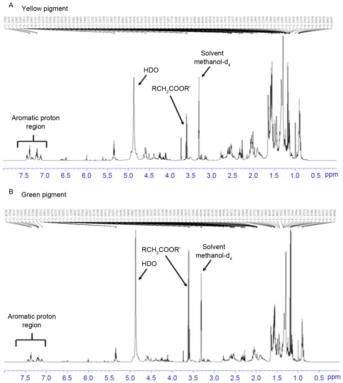
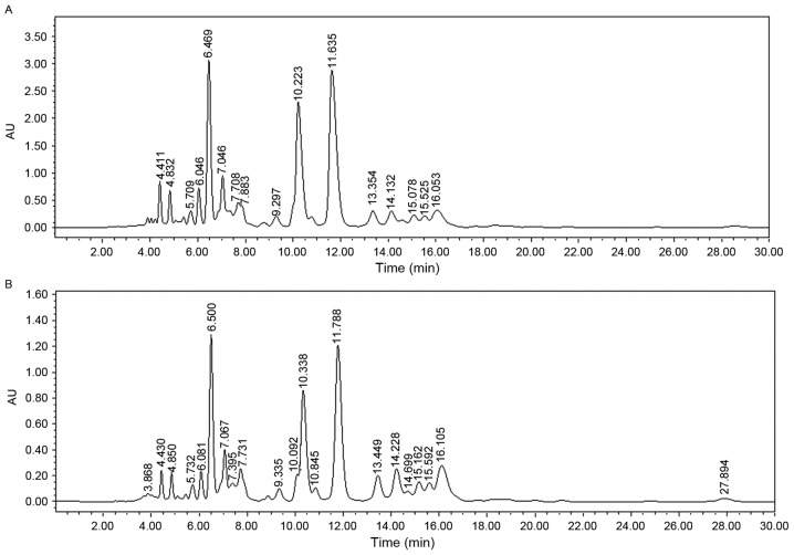
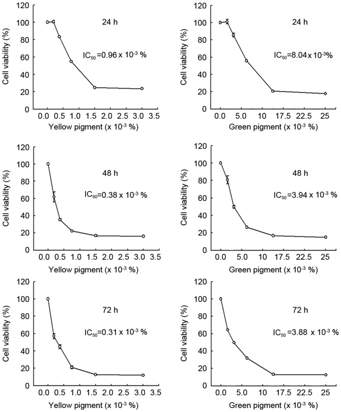
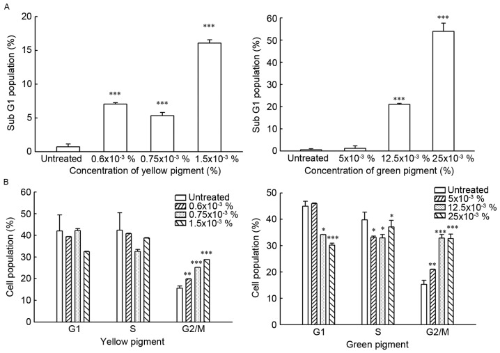
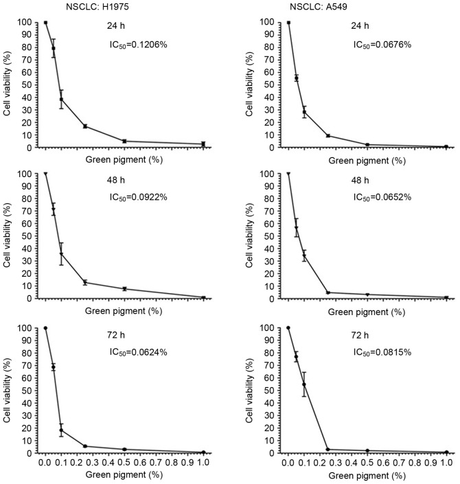
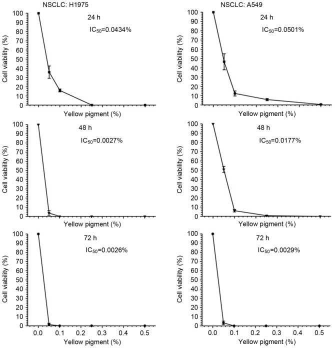
Natural compounds have been candidates for anticancer medicine over the last 20 years. During the process of isolating seed oil from Calophyllum inophyllum L., yellow and green pigments containing multiple compounds with an aromatic structure were identified. High-performance liquid chromatography and nuclear magnetic resonance analysis of these pigments revealed that the compounds present were identical, but the concentration of the compounds was different. Treatment with the pigments was able to induce the death of DLD-1 human colon cancer cells and increase the percentage of the cells in the sub-G1 and sub-G2/M phases in a dose-dependent manner. Additionally, the pigments were able to exhibit cytotoxic activity on A549 and H1975 human non-small cell lung cancer (NSCLC) cell lines at 24 h, with half-maximal inhibitory concentrations (IC50) values of 0.1206 and 0.0676%, respectively for green pigments, and 0.0434 and 0.0501%, respectively for yellow pigments. Furthermore, a decrease in IC50 value was associated with an increase in the duration of treatment. However, a sharp decrease in IC50 value of the yellow pigment was observed for H1975 cells at 48 h and for A549 cells at 72 h compared with no change in IC50 value for the green pigment with time, suggesting that the pigments function and induce cell death differently in the two cell lines. An investigation was performed into the synergistic effect of the green pigment and gefitinib (Iressa®, ZD1839), which is a selective epidermal growth factor receptor-tyrosine kinase inhibitor to block growth factor-mediated cell proliferation. The combination of the green pigment and gefitinib resulted in an enhancement of the decrease in viability of A549 and H1975 cells compared with treatment with gefitinib alone, which suggested that treatment with the green pigments was able to enhance the sensitivity of NSCLC cells to gefitinib. In conclusion, these pigments may be considered for development as anti-colon cancer agents.
Keywords: apoptosis; colon cancer; gefitinib; high-performance liquid chromatography; non-small cell lung cancer; pigments.
Figures
Similar articles
-
Down-regulation of ERK1/2 and AKT-mediated X-ray repair cross-complement group 1 protein (XRCC1) expression by Hsp90 inhibition enhances the gefitinib-induced cytotoxicity in human lung cancer cells.Exp Cell Res. 2015 May 15;334(1):126-35. doi: 10.1016/j.yexcr.2015.01.016. Epub 2015 Feb 4. Exp Cell Res. 2015. PMID: 25662161
-
Gefitinib ("Iressa", ZD1839), an epidermal growth factor receptor tyrosine kinase inhibitor, reverses breast cancer resistance protein/ABCG2-mediated drug resistance.Cancer Res. 2005 Feb 15;65(4):1541-6. doi: 10.1158/0008-5472.CAN-03-2417. Cancer Res. 2005. PMID: 15735043
-
PA-MSHA in combination with EGFR tyrosine kinase inhibitor: A new strategy to overcome the drug resistance of non-small cell lung cancer cells.Oncotarget. 2016 Aug 2;7(31):49384-49396. doi: 10.18632/oncotarget.9891. Oncotarget. 2016. PMID: 27283902 Free PMC article.
-
Anticancer Activity and Molecular Mechanism of Polyphenol Rich Calophyllum inophyllum Fruit Extract in MCF-7 Breast Cancer Cells.Nutr Cancer. 2017 Nov-Dec;69(8):1308-1324. doi: 10.1080/01635581.2017.1367944. Epub 2017 Oct 25. Nutr Cancer. 2017. PMID: 29068745
-
Gefitinib (Iressa, ZD1839): a novel targeted approach for the treatment of solid tumors.Bull Cancer. 2004 May 1;91(5):E70-6. Bull Cancer. 2004. PMID: 15582898 Review.
Cited by
-
Prodigiosin-Emerged PI3K/Beclin-1-Independent Pathway Elicits Autophagic Cell Death in Doxorubicin-Sensitive and -Resistant Lung Cancer.J Clin Med. 2018 Oct 3;7(10):321. doi: 10.3390/jcm7100321. J Clin Med. 2018. PMID: 30282915 Free PMC article.
-
Phytochemical Profiles and Anticancer Effects of Calophyllum inophyllum L. Extract Relating to Reactive Oxygen Species Modulation on Patient-Derived Cells from Breast and Lung Cancers.Scientifica (Cairo). 2023 Jul 20;2023:6613670. doi: 10.1155/2023/6613670. eCollection 2023. Scientifica (Cairo). 2023. PMID: 37520043 Free PMC article.
References
LinkOut - more resources
Full Text Sources
Other Literature Sources
Research Materials
Miscellaneous