

Interrupted aortic arch diagnosis by computed tomography angiography and 3-D reconstruction: A case report
- PMID: 29552241
- PMCID: PMC5851190
- DOI: 10.1016/j.radcr.2017.10.001
Interrupted aortic arch diagnosis by computed tomography angiography and 3-D reconstruction: A case report
Abstract
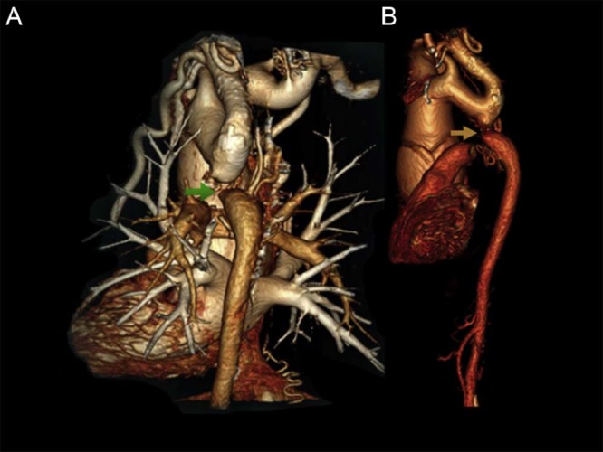
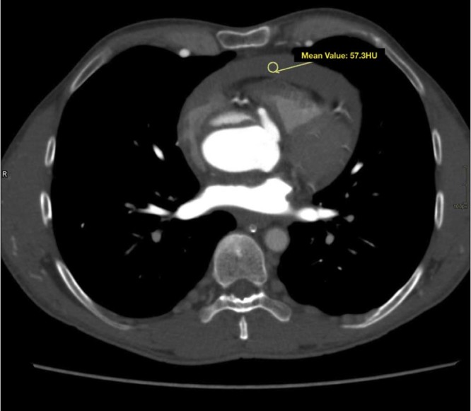
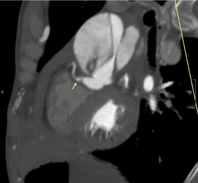
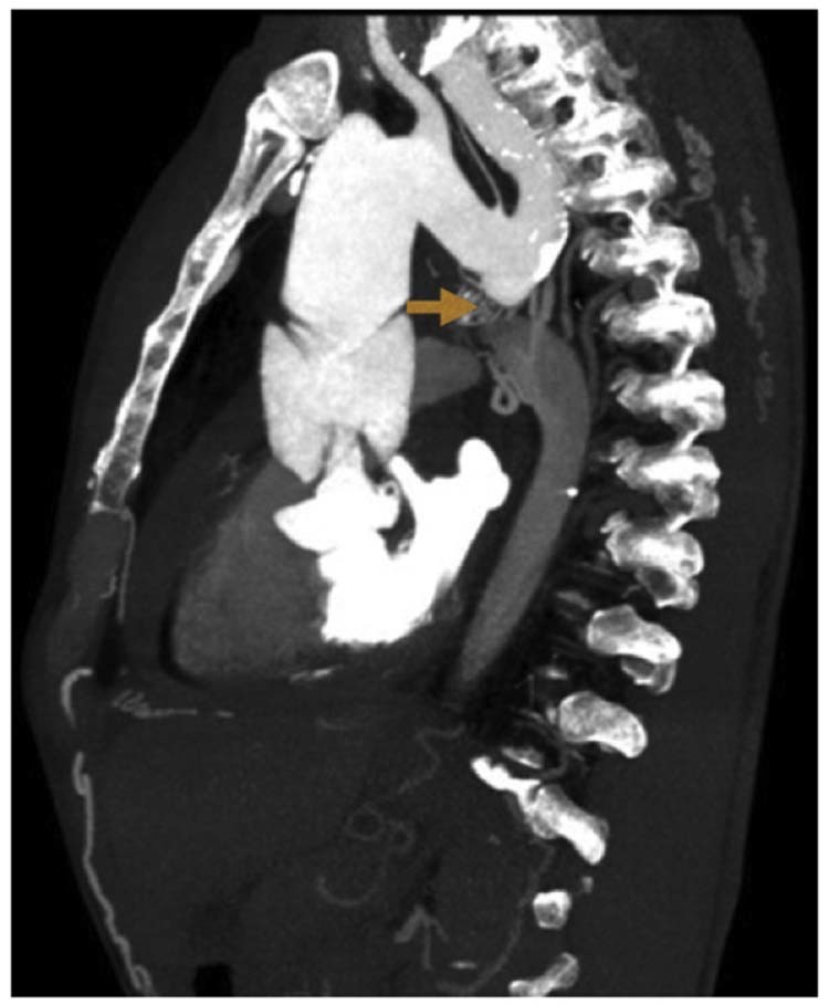
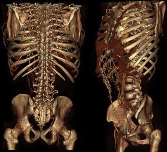
Interrupted aortic arch is an extremely rare congenital malformation representing about 1% of congenital heart disease. Early symptoms usually occur early in the neonatal period and clinical deterioration is often rapid and long-term prognosis is limited. Nonetheless, this condition has been identified later in adult life in rare cases. We report a case in an adult male with absence of hypertension history and no further cardiac compromise, with a severe posterior chest pain alongside dyspnea and sweating. Computed tomography angiography revealed interrupted aortic arch type A, bivalve aorta, hemopericardium, aortic dissection Stanford A, and important collateral circulation.
Keywords: Computed tomography angiography; Diagnostic imaging; Interrupted aortic arch.
Figures
Similar articles
-
Rare case series of adult interrupted aortic arch.J Cardiol Cases. 2023 Jul 29;28(5):206-209. doi: 10.1016/j.jccase.2023.07.004. eCollection 2023 Nov. J Cardiol Cases. 2023. PMID: 38024110 Free PMC article.
-
[Interrupted aortic arch in a 42-year-old man].Nan Fang Yi Ke Da Xue Xue Bao. 2012 Apr;32(4):593-4. Nan Fang Yi Ke Da Xue Xue Bao. 2012. PMID: 22543152 Chinese.
-
Isolated interrupted aortic arch accompanied by type B aortic dissection and extensive collateral arteries diagnosed with MDCT angiography.Clin Imaging. 2012 Sep-Oct;36(5):602-5. doi: 10.1016/j.clinimag.2011.12.021. Epub 2012 Jun 8. Clin Imaging. 2012. PMID: 22920371
-
In Utero Aortic Arch Thrombosis Masquerading as Interrupted Aortic Arch: A Case Report and Review of the Literature.Pediatr Cardiol. 2019 Mar;40(3):658-663. doi: 10.1007/s00246-019-02068-5. Epub 2019 Feb 8. Pediatr Cardiol. 2019. PMID: 30734851 Review.
-
Congenital Variants and Anomalies of the Aortic Arch.Radiographics. 2017 Jan-Feb;37(1):32-51. doi: 10.1148/rg.2017160033. Epub 2016 Nov 18. Radiographics. 2017. PMID: 27860551 Review.
Cited by
-
A comparative analysis of CT angiography and echocardiography in the evaluation of chest findings in patients with interrupted aortic arch.Front Radiol. 2025 Jun 26;5:1616112. doi: 10.3389/fradi.2025.1616112. eCollection 2025. Front Radiol. 2025. PMID: 40642309 Free PMC article.
-
Difference and similarity between type A interrupted aortic arch and aortic coarctation in adults: Two case reports.World J Clin Cases. 2022 Apr 16;10(11):3472-3477. doi: 10.12998/wjcc.v10.i11.3472. World J Clin Cases. 2022. PMID: 35611201 Free PMC article.
References
-
- Dillman J.R., Yarram S.G., D'Amico A.R., Hernandez R.J. Interrupted aortic arch: spectrum of MRI findings. AJR Am J Roentgenol. 2008;190:1467–1474. - PubMed
-
- Gordon E.A., Person T., Kavarana M., Ikonomidis J.S. Interrupted aortic arch in the adult. J Card Surg. 2011;26:405–409. - PubMed
-
- Ponte M., Dias A., Dias Ferreira N., Fonseca C., Mota J.C., Gama V. Interrupted aortic arch: a misdiagnosed cause of hypertension. Rev Port Cardiol. 2014;33:389.e1–389.e5. - PubMed
-
- Erden I., Kayapinar O., Erden E.C., Yalçin S. Silent interrupted aortic arch in an elderly patient. Cardiol J. 2011;18:695–697. - PubMed
Publication types
LinkOut - more resources
Full Text Sources
Other Literature Sources
