

Pediatric Primary Tuberculous Osteomyelitis of the Mandible Mimicking Parotitis
- PMID: 29552433
- PMCID: PMC5854305
- DOI: 10.7759/cureus.2071
Pediatric Primary Tuberculous Osteomyelitis of the Mandible Mimicking Parotitis
Abstract
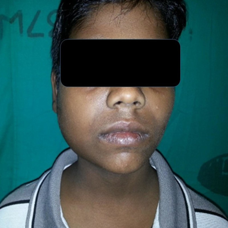
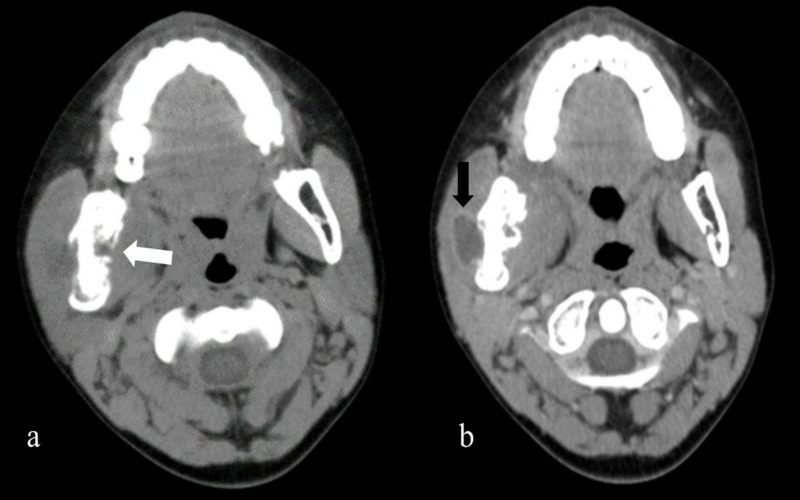
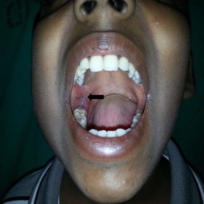
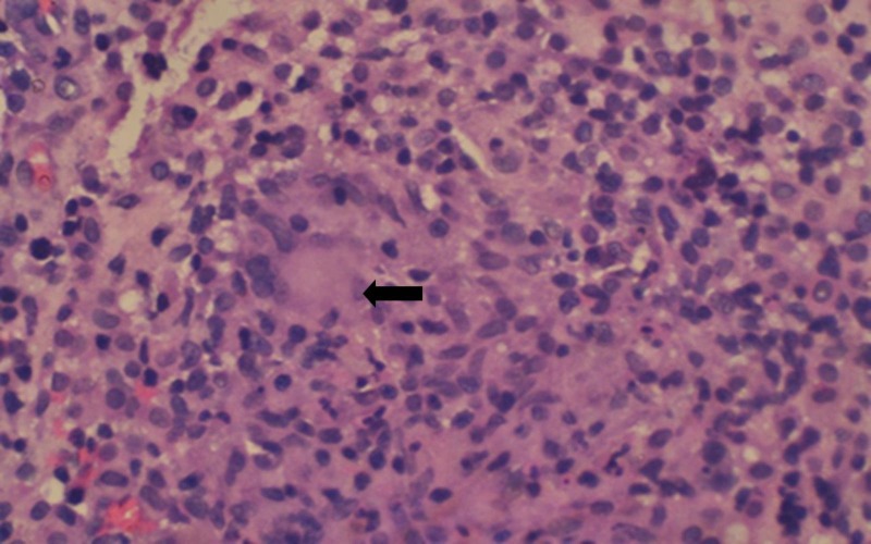
Tuberculosis (TB) is a worldwide public health problem; however, primary tuberculous osteomyelitis involving the mandible is extremely rare. Here, we report a 14-year-old boy who presented with a recurrent, generalized swelling of the cheek in the right side, mimicking parotitis. Fine needle aspiration cytology (FNAC) from the swelling was inconclusive. Contrast-enhanced computed tomography (CECT) of the head and neck revealed an osteolytic lesion of the mandible with a surrounding abscess. An intraoral incisional biopsy of the tissue showed a granulomatous lesion. The patient was started on anti-tubercular therapy (ATT) for six months. Our patient's presentation underscores the clinical difficulty in establishing a diagnosis and considering tuberculous osteomyelitis in the differential diagnosis.
Keywords: lymphadenitis; mandible; osteomyelitis; parotitis; tuberculosis.
Conflict of interest statement
The authors have declared that no competing interests exist.
Figures
Similar articles
-
Primary Mandibular Tuberculous Osteomyelitis Mimicking Ameloblastoma: A Case Report and Literature Review of Mandibular Tuberculous Osteomyelitis.Arch Plast Surg. 2024 Feb 29;51(2):187-195. doi: 10.1055/a-2217-8784. eCollection 2024 Mar. Arch Plast Surg. 2024. PMID: 38596155 Free PMC article.
-
A Case of Primary Tuberculous Parotitis Mimicking Parotid Neoplasm: A Rare Clinical Entity.Cureus. 2024 Apr 13;16(4):e58217. doi: 10.7759/cureus.58217. eCollection 2024 Apr. Cureus. 2024. PMID: 38745804 Free PMC article.
-
Primary tuberculous osteomyelitis of the mandible: a case report.Dentomaxillofac Radiol. 2008 Oct;37(7):415-20. doi: 10.1259/dmfr/73393014. Dentomaxillofac Radiol. 2008. PMID: 18812606
-
Tuberculous Lymphadenitis and Parotitis.Microbiol Spectr. 2016 Dec;4(6). doi: 10.1128/microbiolspec.TNMI7-0008-2016. Microbiol Spectr. 2016. PMID: 28084205 Review.
-
Tuberculous parotitis: case report and literature review.Ann Otol Rhinol Laryngol. 2005 Jul;114(7):547-51. doi: 10.1177/000348940511400710. Ann Otol Rhinol Laryngol. 2005. PMID: 16134352 Review.
Cited by
-
Primary Mandibular Tuberculous Osteomyelitis Mimicking Ameloblastoma: A Case Report and Literature Review of Mandibular Tuberculous Osteomyelitis.Arch Plast Surg. 2024 Feb 29;51(2):187-195. doi: 10.1055/a-2217-8784. eCollection 2024 Mar. Arch Plast Surg. 2024. PMID: 38596155 Free PMC article.
-
Periostitis Ossificans: Report of Two Cases Resolved with Endodontic Treatment.Case Rep Dent. 2020 Nov 24;2020:8876268. doi: 10.1155/2020/8876268. eCollection 2020. Case Rep Dent. 2020. PMID: 33299618 Free PMC article.
-
Juvenile Tuberculous Osteomyelitis of the Mandible: A Rarity.J Maxillofac Oral Surg. 2025 Feb;24(1):70-73. doi: 10.1007/s12663-024-02216-2. Epub 2024 May 31. J Maxillofac Oral Surg. 2025. PMID: 39902452
References
-
- Primary tuberculous osteomyelitis of the mandible. Imamura M, Kakihara T, Yamamoto K, Imai C, Tanaka A, Uchiyama M. Pediatr Int. 2004;46:736–739. - PubMed
-
- Tuberculous osteomyelitis of the mandible: a case report. Bhatt AP, Jayakrishnan A. Int J Paediatr Dent. 2001;11:304–308. - PubMed
-
- Tuberculous osteomyelitis of the mandible with diffuse swelling of the floor of the mouth: a case report. Bai S, Sun CF. J Oral Maxillofac Surg. 2014;72:749. - PubMed
-
- Primary tuberculosis of mandible. Gupta MK, Singh M. http://www.indianpediatrics.net/jan2007/53.pdf. Indian Pediatr. 2007;44:53–54. - PubMed
-
- Primary tuberculous osteomyelitis of the mandible. A case report. Fukuda J, Shingo Y, Miyako H. Oral Surg Oral Med Oral Pathol. 1992;73:278–280. - PubMed
Publication types
LinkOut - more resources
Full Text Sources
Other Literature Sources