

Network connectivity of motor control in the ageing brain
- PMID: 29552486
- PMCID: PMC5852391
- DOI: 10.1016/j.nicl.2018.02.001
Network connectivity of motor control in the ageing brain
Abstract
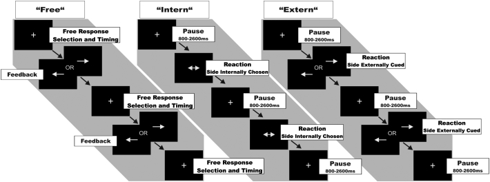
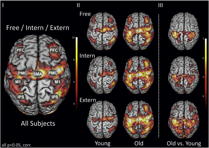
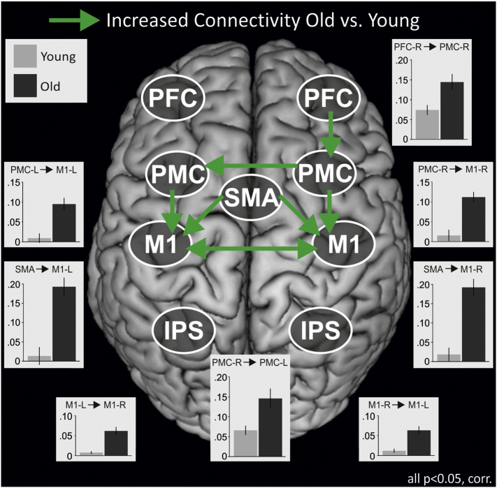
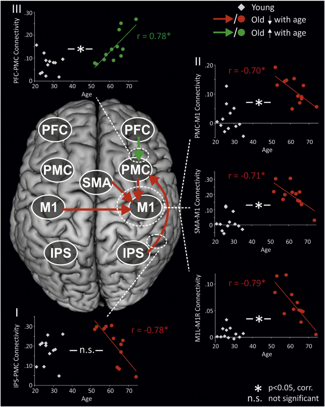
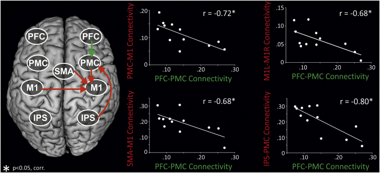
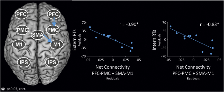
Older individuals typically display stronger regional brain activity than younger subjects during motor performance. However, knowledge regarding age-related changes of motor network interactions between brain regions remains scarce. We here investigated the impact of ageing on the interaction of cortical areas during movement selection and initiation using dynamic causal modelling (DCM). We found that age-related psychomotor slowing was accompanied by increases in both regional activity and effective connectivity, especially for 'core' motor coupling targeting primary motor cortex (M1). Interestingly, younger participants within the older group showed strongest connectivity targeting M1, which steadily decreased with advancing age. Conversely, prefrontal influences on the motor system increased with advancing age, and were inversely correlated with reduced parietal influences and core motor coupling. Interestingly, higher net coupling within the prefrontal-premotor-M1 axis predicted faster psychomotor speed in ageing. Hence, as opposed to a uniform age-related decline, our findings are compatible with the idea of different age-related compensatory mechanisms, with an important role of the prefrontal cortex compensating for reduced coupling within the core motor network.
Keywords: Ageing; Effective connectivity; Motor control; fMRI.
Figures
Similar articles
-
Dopaminergic modulation of motor network dynamics in Parkinson's disease.Brain. 2015 Mar;138(Pt 3):664-78. doi: 10.1093/brain/awu381. Epub 2015 Jan 6. Brain. 2015. PMID: 25567321 Free PMC article.
-
Ageing changes effective connectivity of motor networks during bimanual finger coordination.Neuroimage. 2016 Dec;143:325-342. doi: 10.1016/j.neuroimage.2016.09.014. Epub 2016 Sep 9. Neuroimage. 2016. PMID: 27616642
-
Directed connectivity between primary and premotor areas underlying ankle force control in young and older adults.Neuroimage. 2020 Sep;218:116982. doi: 10.1016/j.neuroimage.2020.116982. Epub 2020 May 22. Neuroimage. 2020. PMID: 32450250
-
Dynamic causal modelling of EEG and fMRI to characterize network architectures in a simple motor task.Neuroimage. 2016 Jan 1;124(Pt A):498-508. doi: 10.1016/j.neuroimage.2015.08.052. Epub 2015 Aug 31. Neuroimage. 2016. PMID: 26334836
-
Motor control and aging: links to age-related brain structural, functional, and biochemical effects.Neurosci Biobehav Rev. 2010 Apr;34(5):721-33. doi: 10.1016/j.neubiorev.2009.10.005. Epub 2009 Oct 20. Neurosci Biobehav Rev. 2010. PMID: 19850077 Free PMC article. Review.
Cited by
-
Differential Cortical and Subcortical Activations during Different Stages of Muscle Control: A Functional Magnetic Resonance Imaging Study.Brain Sci. 2024 Apr 20;14(4):404. doi: 10.3390/brainsci14040404. Brain Sci. 2024. PMID: 38672052 Free PMC article.
-
Investigating the effects of age and conditioning stimulation intensity on SMA-M1 connectivity in younger, middle-aged, and older adults.Eur J Appl Physiol. 2025 Jul 21. doi: 10.1007/s00421-025-05904-0. Online ahead of print. Eur J Appl Physiol. 2025. PMID: 40689955
-
Primary motor cortical activity during unimanual movements with increasing demand on precision.J Neurophysiol. 2020 Sep 1;124(3):728-739. doi: 10.1152/jn.00546.2019. Epub 2020 Jul 29. J Neurophysiol. 2020. PMID: 32727264 Free PMC article.
-
An fMRI Study of the Brain Network Involved in Teeth Tapping in Elderly Adults.Front Aging Neurosci. 2020 Mar 17;12:32. doi: 10.3389/fnagi.2020.00032. eCollection 2020. Front Aging Neurosci. 2020. PMID: 32256334 Free PMC article.
-
A distributed theta network of error generation and processing in aging.Cogn Neurodyn. 2024 Apr;18(2):447-459. doi: 10.1007/s11571-023-10018-4. Epub 2023 Nov 16. Cogn Neurodyn. 2024. PMID: 38699606 Free PMC article.
References
-
- Ankudowich E., Pasvanis S., Rajah M.N. Changes in the correlation between spatial and temporal source memory performance and BOLD activity across the adult lifespan. Cortex. 2017;91:234–249. - PubMed
-
- Ashburner J., Friston K.J. Unified segmentation. NeuroImage. 2005;26:839–851. - PubMed
-
- Bates J.F., Goldman-Rakic P.S. Prefrontal connections of medial motor areas in the rhesus monkey. J. Comp. Neurol. 1993;336:211–228. - PubMed
-
- Benjamini Y., Hochberg Y. Controlling the false discovery rate: a practical and powerful approach to multiple testing. J. R. Stat. Soc. Ser. B (Methodol.) 1995;57:289–300.
-
- Berchicci M., Lucci G., Pesce C., Spinelli D., Di Russo F. Prefrontal hyperactivity in older people during motor planning. NeuroImage. 2012;62:1750–1760. - PubMed
Publication types
MeSH terms
Substances
Grants and funding
LinkOut - more resources
Full Text Sources
Other Literature Sources
Medical
