Review
. 2018 Mar 2:8:61.
doi: 10.3389/fcimb.2018.00061. eCollection 2018.

Acanthamoeba and Dictyostelium as Cellular Models for Legionella Infection

Affiliations
Review

Acanthamoeba and Dictyostelium as Cellular Models for Legionella Infection

A Leoni Swart et al. Front Cell Infect Microbiol. .

Abstract

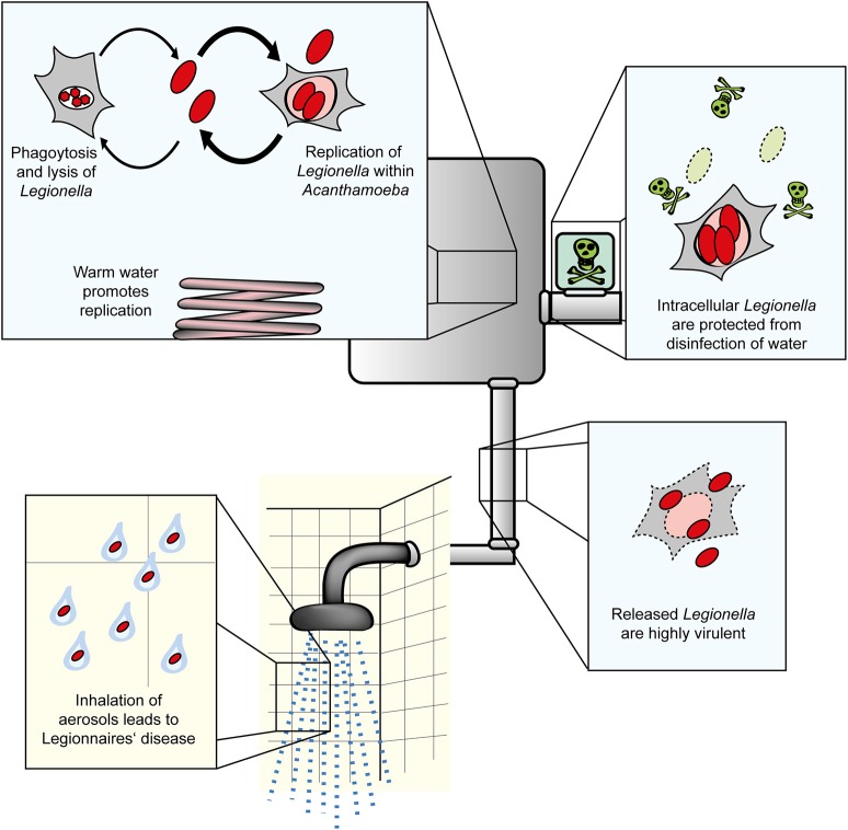
Environmental bacteria of the genus Legionella naturally parasitize free-living amoebae. Upon inhalation of bacteria-laden aerosols, the opportunistic pathogens grow intracellularly in alveolar macrophages and can cause a life-threatening pneumonia termed Legionnaires' disease. Intracellular replication in amoebae and macrophages takes place in a unique membrane-bound compartment, the Legionella-containing vacuole (LCV). LCV formation requires the bacterial Icm/Dot type IV secretion system, which translocates literally hundreds of "effector" proteins into host cells, where they modulate crucial cellular processes for the pathogen's benefit. The mechanism of LCV formation appears to be evolutionarily conserved, and therefore, amoebae are not only ecologically significant niches for Legionella spp., but also useful cellular models for eukaryotic phagocytes. In particular, Acanthamoeba castellanii and Dictyostelium discoideum emerged over the last years as versatile and powerful models. Using genetic, biochemical and cell biological approaches, molecular interactions between amoebae and Legionella pneumophila have recently been investigated in detail with a focus on the role of phosphoinositide lipids, small and large GTPases, autophagy components and the retromer complex, as well as on bacterial effectors targeting these host factors.

Keywords: GTPase; amoebae; effector protein; host-pathogen interaction; pathogen vacuole; phosphoinositide lipid; retrograde transport; type IV secretion.


Figures

Figure 1
Figure 1
Amoebae as natural reservoir for Legionella in water systems. L. pneumophila and amoebae are commonly isolated from natural and technical water systems. At cooler temperatures (below ca. 20–25°C), L. pneumophila resists A. castellanii, but does not or only slowly replicate in the amoebae. At elevated temperatures occurring in technical water systems, L. pneumophila efficiently replicates in the amoebae. Internalized Legionella bacteria are less susceptible to routine disinfection and released from the amoebae in a highly virulent form. Virulent bacteria present in aerosols are inhaled, infect alveolar macrophages and cause Legionnaires' disease.
Figure 2
Figure 2
Experimental approaches to study Legionella infection of amoebae. (A) Amoeba plate test showing serial dilutions of bacterial cultures spotted on a lawn of A. castellanii. Virulent L. pneumophila is amoeba-resistant and grows in high dilutions, while mutant strains lacking a functional Icm/Dot T4SS do not (ΔicmT) or barely (ΔicmS) grow. Growth defect of the mutants is complemented by plasmid-borne expression of the corresponding genes (ΔicmT+icmT, ΔicmS+icmS). (B) Intracellular replication quantified by fluorescence from GFP-producing L. pneumophila. Virulent bacteria show an increase in fluorescence over time, while ΔicmT mutant bacteria do not grow. (C) Fluorescence microscopy images of L. pneumophila-infected, dying amoebae that round up, losing the characteristic spiky morphology, and take up the fluorescent dye propidium iodide. (D) Live-cell confocal fluorescence microscopy of D. discoideum strain Ax3 producing P4C-mCherry (red) and GFP-Sey1 (green), infected (MOI 10, 2 h) with mCerulean-producing L. pneumophila JR32 (white). Scale bar: 2 μm. (E) Flow cytometry of A. castellanii infected with L. pneumophila wild-type or ΔicmT and stained with propidium iodide. Dye uptake and changes in morphology are quantified by fluorescence and light scattering, respectively. (F) Flow cytometry gating on infected A. castellanii reveals replication of GFP-producing intracellular L. pneumophila wild-type but not ΔicmT mutant bacteria. (G) LCV isolation by immuno-magnetic separation and density gradient centrifugation from lysates of D. discoideum Ax3, infected (MOI 10, 2 h) with DsRed-producing L. pneumophila JR32 (red). The preparation was immuno-stained with an anti-calnexin antibody, followed by a FITC-coupled secondary antibody (green). Scale bar: 10 μm, zoom: 2 μm. Image (A) was reproduced with permission from Albers et al. (2005) and images (C,E,F) from Tiaden et al. (2007).

References

    1. Albers U., Reus K., Shuman H. A., Hilbi H. (2005). The amoebae plate test implicates a paralogue of lpxB in the interaction of Legionella pneumophila with Acanthamoeba castellanii. Microbiology 151, 167–182. 10.1099/mic.0.27563-0 - DOI - PubMed
    1. Albers U., Tiaden A., Spirig T., Al Alam D., Goyert S. M., Gangloff S. C., et al. . (2007). Expression of Legionella pneumophila paralogous lipid A biosynthesis genes under different growth conditions. Microbiology 153, 3817–3829. 10.1099/mic.0.2007/009829-0 - DOI - PubMed
    1. Al-Quadan T., Kwaik Y. A. (2011). Molecular characterization of exploitation of the polyubiquitination and farnesylation machineries of Dictyostelium discoideum by the AnkB F-Box effector of Legionella pneumophila. Front. Microbiol. 2:23. 10.3389/fmicb.2011.00023 - DOI - PMC - PubMed
    1. Amaro F., Wang W., Gilbert J. A., Anderson O. R., Shuman H. A. (2015). Diverse protist grazers select for virulence-related traits in Legionella. ISME J. 9, 1607–1618. 10.1038/ismej.2014.248 - DOI - PMC - PubMed
    1. Asrat S., de Jesús D. A., Hempstead A. D., Ramabhadran V., Isberg R. R. (2014). Bacterial pathogen manipulation of host membrane trafficking. Annu. Rev. Cell Dev. Biol. 30, 79–109. 10.1146/annurev-cellbio-100913-013439 - DOI - PubMed

Publication types

MeSH terms

LinkOut - more resources