

Target-Cell-Directed Bioengineering Approaches for Gene Therapy of Hemophilia A
- PMID: 29552578
- PMCID: PMC5852392
- DOI: 10.1016/j.omtm.2018.01.004
Target-Cell-Directed Bioengineering Approaches for Gene Therapy of Hemophilia A
Abstract
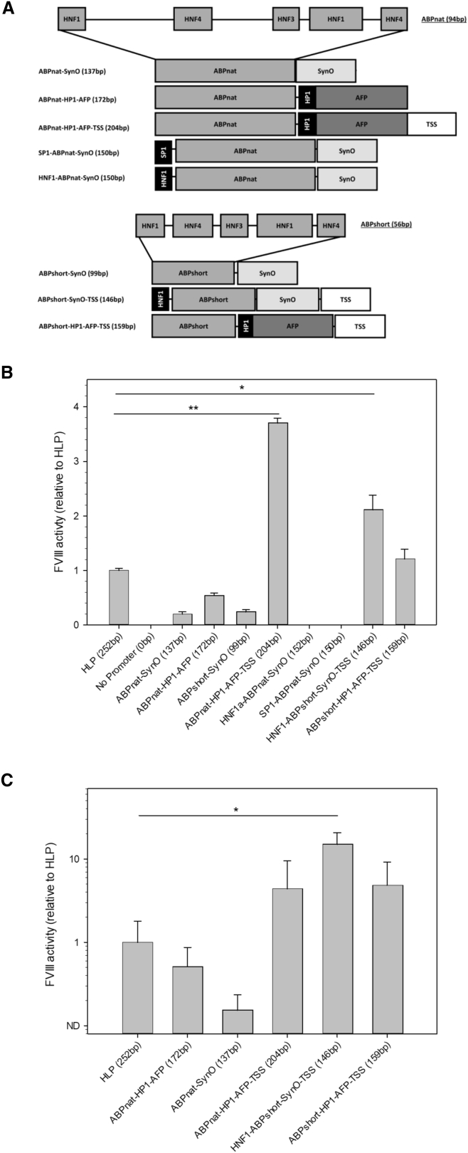
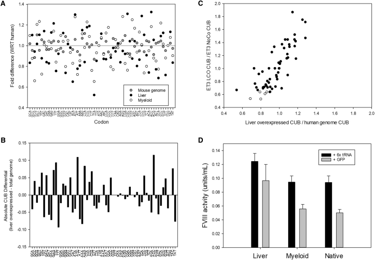
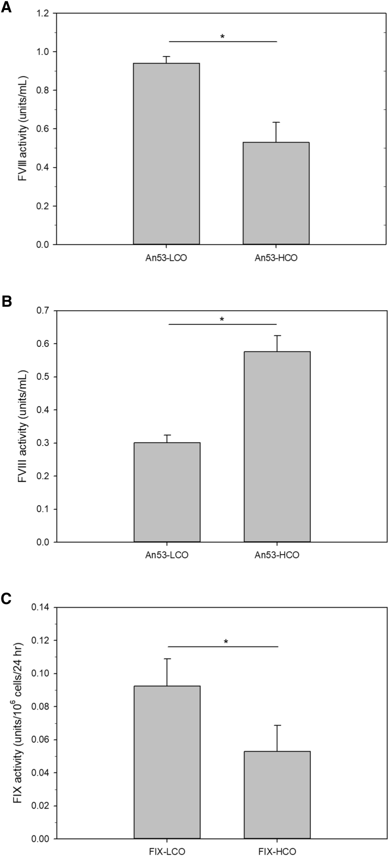
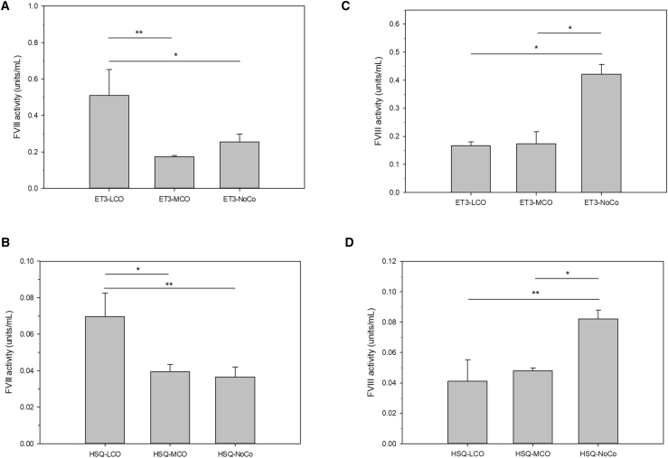
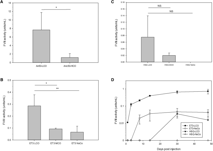
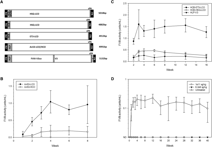
Potency is a key optimization parameter for hemophilia A gene therapy product candidates. Optimization strategies include promoter engineering to increase transcription, codon optimization of mRNA to improve translation, and amino-acid substitution to promote secretion. Herein, we describe both rational and empirical design approaches to the development of a minimally sized, highly potent AAV-fVIII vector that incorporates three unique elements: a liver-directed 146-nt transcription regulatory module, a target-cell-specific codon optimization algorithm, and a high-expression bioengineered fVIII variant. The minimal synthetic promoter allows for the smallest AAV-fVIII vector genome known at 4,832 nt, while the tissue-directed codon optimization strategy facilitates increased fVIII transgene product expression in target cell types, e.g., hepatocytes, over traditional genome-level codon optimization strategies. As a tertiary approach, we incorporated ancient and orthologous fVIII sequence elements previously shown to facilitate improved biosynthesis through post-translational mechanisms. Together, these technologies contribute to an AAV-fVIII vector that confers sustained, curative levels of fVIII at a minimal dose in hemophilia A mice. Moreover, the first two technologies should be generalizable to all liver-directed gene therapy vector designs.
Keywords: AAV; codon optimization; factor VIII; hemophilia; promoter design; vector optimization.
Figures
Similar articles
-
Bioengineered coagulation factor VIII enables long-term correction of murine hemophilia A following liver-directed adeno-associated viral vector delivery.Mol Ther Methods Clin Dev. 2014 Aug 6;1:14036. doi: 10.1038/mtm.2014.36. eCollection 2014. Mol Ther Methods Clin Dev. 2014. PMID: 26015976 Free PMC article.
-
Preclinical assessment of an optimized AAV-FVIII vector in mice and non-human primates for the treatment of hemophilia A.Mol Ther Methods Clin Dev. 2021 Nov 24;24:20-29. doi: 10.1016/j.omtm.2021.11.005. eCollection 2022 Mar 10. Mol Ther Methods Clin Dev. 2021. PMID: 34977269 Free PMC article.
-
Effects of FVIII immunity on hepatocyte and hematopoietic stem cell-directed gene therapy of murine hemophilia A.Mol Ther Methods Clin Dev. 2016 Feb 10;3:15056. doi: 10.1038/mtm.2015.56. eCollection 2016. Mol Ther Methods Clin Dev. 2016. PMID: 26909355 Free PMC article.
-
Hemophilia Gene Therapy: Ready for Prime Time?Hum Gene Ther. 2017 Nov;28(11):1013-1023. doi: 10.1089/hum.2017.116. Epub 2017 Aug 3. Hum Gene Ther. 2017. PMID: 28793786 Review.
-
Hemophilia A gene therapy via intraosseous delivery of factor VIII-lentiviral vectors.Thromb J. 2016 Oct 4;14(Suppl 1):41. doi: 10.1186/s12959-016-0105-1. eCollection 2016. Thromb J. 2016. PMID: 27766066 Free PMC article. Review.
Cited by
-
Thrombopoietin-based CAR-T cells demonstrate in vitro and in vivo cytotoxicity to MPL positive acute myelogenous leukemia and hematopoietic stem cells.Gene Ther. 2022 May;29(5):1-12. doi: 10.1038/s41434-021-00283-5. Epub 2021 Aug 13. Gene Ther. 2022. PMID: 34385604
-
Preclinical development of TAK-754, a high-performance AAV8-based vector expressing coagulation factor VIII.Mol Ther Methods Clin Dev. 2025 Jan 28;33(1):101424. doi: 10.1016/j.omtm.2025.101424. eCollection 2025 Mar 13. Mol Ther Methods Clin Dev. 2025. PMID: 40123744 Free PMC article.
-
Transplanting FVIII/ET3-secreting cells in fetal sheep increases FVIII levels long-term without inducing immunity or toxicity.Nat Commun. 2023 Jul 14;14(1):4206. doi: 10.1038/s41467-023-39986-1. Nat Commun. 2023. PMID: 37452013 Free PMC article.
-
The Immune Response to the fVIII Gene Therapy in Preclinical Models.Front Immunol. 2020 Apr 15;11:494. doi: 10.3389/fimmu.2020.00494. eCollection 2020. Front Immunol. 2020. PMID: 32351497 Free PMC article. Review.
-
Enhancing the effectiveness of γδ T cells by mRNA transfection of chimeric antigen receptors or bispecific T cell engagers.Mol Ther Oncolytics. 2023 May 22;29:145-157. doi: 10.1016/j.omto.2023.05.007. eCollection 2023 Jun 15. Mol Ther Oncolytics. 2023. PMID: 37387794 Free PMC article.
References
-
- Kay M.A., Manno C.S., Ragni M.V., Larson P.J., Couto L.B., McClelland A., Glader B., Chew A.J., Tai S.J., Herzog R.W. Evidence for gene transfer and expression of factor IX in haemophilia B patients treated with an AAV vector. Nat. Genet. 2000;24:257–261. - PubMed
-
- Manno C.S., Pierce G.F., Arruda V.R., Glader B., Ragni M., Rasko J.J., Ozelo M.C., Hoots K., Blatt P., Konkle B. Successful transduction of liver in hemophilia by AAV-Factor IX and limitations imposed by the host immune response. Nat. Med. 2006;12:342–347. - PubMed
-
- Nathwani A.C., Gray J.T., Ng C.Y., Zhou J., Spence Y., Waddington S.N., Tuddenham E.G., Kemball-Cook G., McIntosh J., Boon-Spijker M. Self-complementary adeno-associated virus vectors containing a novel liver-specific human factor IX expression cassette enable highly efficient transduction of murine and nonhuman primate liver. Blood. 2006;107:2653–2661. - PMC - PubMed
Grants and funding
LinkOut - more resources
Full Text Sources
Other Literature Sources
Research Materials
