. 2018 Jan 31:9:81-89.
doi: 10.1016/j.omtm.2018.01.007. eCollection 2018 Jun 15.

Plastin 3 Promotes Motor Neuron Axonal Growth and Extends Survival in a Mouse Model of Spinal Muscular Atrophy

Affiliations

Plastin 3 Promotes Motor Neuron Axonal Growth and Extends Survival in a Mouse Model of Spinal Muscular Atrophy

Aziza Alrafiah et al. Mol Ther Methods Clin Dev. .

Abstract

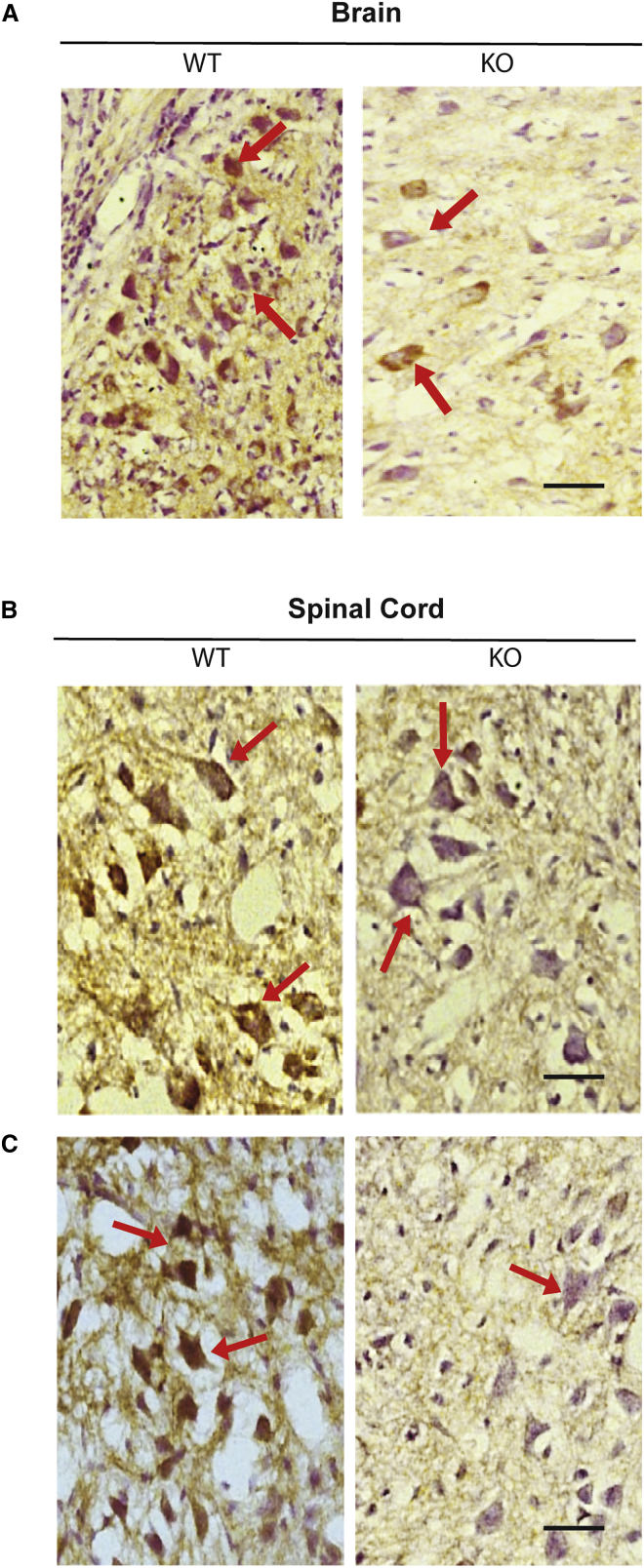
Spinal muscular atrophy (SMA) is a devastating childhood motor neuron disease. SMA is caused by mutations in the survival motor neuron gene (SMN1), leading to reduced levels of SMN protein in the CNS. The actin-binding protein plastin 3 (PLS3) has been reported as a modifier for SMA, making it a potential therapeutic target. Here, we show reduced levels of PLS3 protein in the brain and spinal cord of a mouse model of SMA. Our study also revealed that lentiviral-mediated PLS3 expression restored axonal length in cultured Smn-deficient motor neurons. Delivery of adeno-associated virus serotype 9 (AAV9) harboring Pls3 cDNA via cisterna magna in SMNΔ7 mice, a widely used animal model of SMA, led to high neuronal transduction efficiency. PLS3 treatment allowed a small but significant increase of lifespan by 42%. Although there was no improvement of phenotype, this study has demonstrated the potential use of Pls3 as a target for gene therapy, possibly in combination with other disease modifiers.

Keywords: SMA; gene therapy; plastin 3.


Figures

Figure 1
Figure 1
Plastin 3 Protein Levels in Postnatal Day 1, 5, and 10 SMNΔ7 Mice Representative images of western blot and fold changes in PLS3 protein levels in brain, spinal cord, and muscle tissues from wild-type (WT), carrier littermates (HEMI), and SMNΔ7 (KO) postnatal day 1 (P1) (A and B), P5 (C and D), and P10 (E and F) neonate mouse pups (n = 3). The membrane was probed with mouse anti-PLS3 antibody; GAPDH was used as the house keeping control (n = 3). Error bars represent SEM. One-way ANOVA *p < 0.05, **p < 0.01, ***p < 0.001.
Figure 2
Figure 2
PLS3 Immunoreactivity in the Brain and Spinal Cord Sections of WT and KO P10 Pups (A) Immunohistochemical staining showing PLS3 expression in the brain from P10 wild-type (WT) and SMNΔ7 mice (KO) (n = 3 per group). Brain sections from WT showing immune reactivity of PLS3 associated with a large number of neurons in the brain cortex (brown) (arrows). SMNΔ7 mice (KO) brain sections showed reduced reactivity of PLS3 associated with neurons (arrows). Scale bar, 50 μm. PLS3 immunoreactivity in the cervical (B) and lumbar (C) spinal cord from P10 wild-type (WT) and SMNΔ7 (KO) mice (n = 3). WT spinal cord sections showing PLS3 immuno-reactivity in large number of neurons in the ventral horn of the spinal cord (brown) (arrows), when compared to sections from SMNΔ7 mice KO section, which showed minimal reactivity of PLS3 associated with neurons (arrows). Scale bar, 50 μm.
Figure 3
Figure 3
Lentiviral-Mediated Pls3 Expression Rescues Axonal Deficit in Smn-Deficient Motor Neurons (A) Western blot showing NSC34 cells transduced with LV-PLS3 at MOI 5, 10, 20, 40, or 80 and probed with anti-human PLS3 antibody; α-tubulin was the loading control (n = 3). Untransduced (UT) cells were considered as negative control. (B) A dose-response curve of LV-Pls3 at 5 days posttransduction, along with fold change in Pls3 protein levels for each MOI used. (C) Motor neurons isolated from E13 wild-type (WT), carrier (HEMI), and SMNΔ7 (KO) embryos were transduced with LV-Pls3 or LV-GFP at MOI 80. Cells were labeled for Pls3 (blue), tubulin (red), or GFP (green). Scale bars, 20 μm. (D) Impact of LV-Pls3 treatment on axonal length in Smn-deficient motor neurons. The bar chart shows the effect of LV-Pls3 treatment on axonal length in wild-type, carrier (HEMI), and SMN knockout (KO) embryos. One-way ANOVA (*p < 0.05, ***p < 0.001) was considered to be statistically significant. Data generated from four independent experiments (n = 4). Average neurite lengths ± SEM (n = 100 neurites).
Figure 4
Figure 4
AAV9-Mediated Gene Therapy in SMA Mouse Model (A) Western blot analysis of the spinal cord following cisterna magna delivery of AAV9-Pls3. PLS3 protein levels in three parts of the spinal cord; cervical (C), thoracic (T), and lumbar (L) of carrier (HEMI) pups 4 weeks postdelivery of AAV9-GFP or AAV9-Pls3. The membrane was incubated with anti-PLS3 antibody. (B) Body weight growth in SMNΔ7 injected with AAV9-Pls3 (n = 7), AAV9-GFP (n = 7), or PBS controls (n = 7). *p < 0.05, **p < 0.01, ***p < 0.001, ****p < 0.0001; AAV9-Pls3 versus AAV9-GFP; two-way ANOVA with Dunnett’s multiple comparisons test. (C) Kaplan-Meier survival plot to compare lifespan between all experimental groups of mice (n = 7 per group; overall log-rank test p < 0.05 between all three groups; pairwise comparisons with Bonferroni correction *p < 0.05 between PLS3 and PBS and #p < 0.05 between PLS3 and GFP). (D) AAV9-Pls3 transduction efficiency in spinal motor neurons. Carrier littermate pups were injected at postnatal day 1 with AAV9-Pls3; spinal cords were extracted at 4 weeks postinjection. The cervical and lumbar spinal cord sections were fixed and labeled with calcitonin gene-related peptide (CGRP) antibody as a motor neuronal marker (red), PLS3 antibody as an indicator of transduction (green), and DAPI for visualization of nuclei. Scale bar, 20 μm.

References

    1. Oprea G.E., Kröber S., McWhorter M.L., Rossoll W., Müller S., Krawczak M., Bassell G.J., Beattie C.E., Wirth B. Plastin 3 is a protective modifier of autosomal recessive spinal muscular atrophy. Science. 2008;320:524–527. - PMC - PubMed
    1. Stratigopoulos G., Lanzano P., Deng L., Guo J., Kaufmann P., Darras B., Finkel R., Tawil R., McDermott M.P., Martens W. Association of plastin 3 expression with disease severity in spinal muscular atrophy only in postpubertal females. Arch. Neurol. 2010;67:1252–1256. - PubMed
    1. Dimitriadi M., Sleigh J.N., Walker A., Chang H.C., Sen A., Kalloo G., Harris J., Barsby T., Walsh M.B., Satterlee J.S. Conserved genes act as modifiers of invertebrate SMN loss of function defects. PLoS Genet. 2010;6:e1001172. - PMC - PubMed
    1. Lyon A.N., Pineda R.H., Hao T., Kudryashova E., Kudryashov D.S., Beattie C.E. Calcium binding is essential for plastin 3 function in Smn-deficient motoneurons. Hum. Mol. Genet. 2014;23:1990–2004. - PMC - PubMed
    1. McGovern V.L., Gavrilina T.O., Beattie C.E., Burghes A.H. Embryonic motor axon development in the severe SMA mouse. Hum. Mol. Genet. 2008;17:2900–2909. - PMC - PubMed