

The RiboPuromycylation Method (RPM): an Immunofluorescence Technique to Map Translation Sites at the Sub-cellular Level
- PMID: 29552591
- PMCID: PMC5856242
- DOI: 10.21769/BioProtoc.2669
The RiboPuromycylation Method (RPM): an Immunofluorescence Technique to Map Translation Sites at the Sub-cellular Level
Abstract
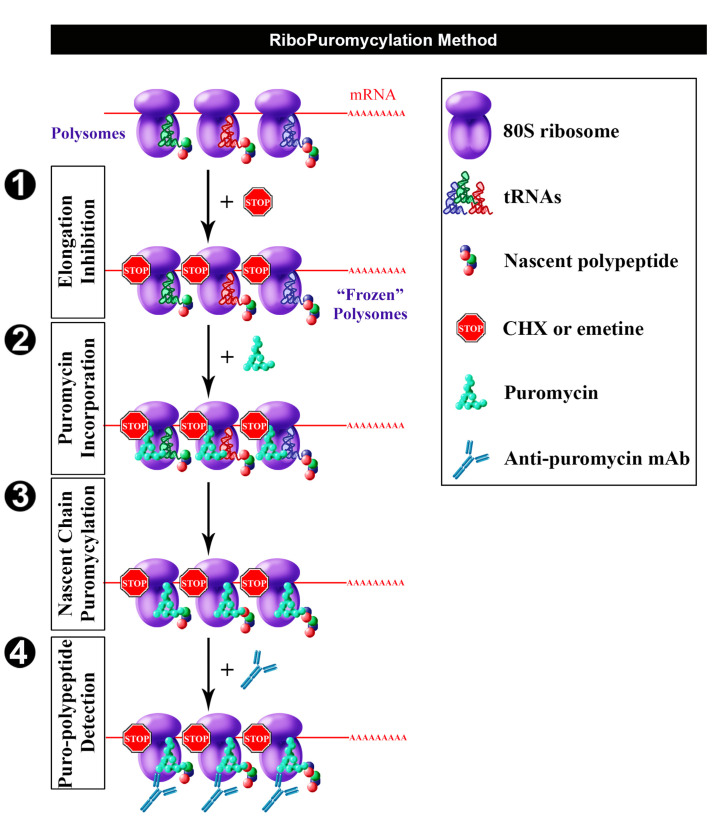
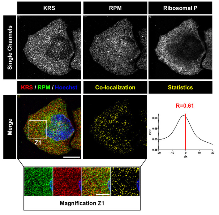
While isotopic labeling of amino acids remains the reference method in the field for quantifying translation rate, it does not provide any information on spatial localization of translation sites. The rationale behind developing the ribopuromycylation method (RPM) was primarily to map translation sites at the sub-cellular level while avoiding detection of newly synthesized proteins released from ribosomes. RPM visualizes actively translating ribosomes in cells via standard immunofluorescence microscopy in fixed and permeabilized cells using a puromycin-specific monoclonal antibody to detect puromycylated nascent chains trapped on ribosomes treated with a chain elongation inhibitor.
Keywords: Nascent chain; Puromycin; Ribopuromycylation; Ribosome; Translation site.
Conflict of interest statement
The authors declare no conflicts of interest or competing interests.
Figures
References
-
- Biever A., Puighermanal E., Nishi A., David A., Panciatici C., Longueville S., Xirodimas D., Gangarossa G., Meyuhas O., Herve D., Girault J. A. and Valjent E.(2015). PKA-dependent phosphorylation of ribosomal protein S6 does not correlate with translation efficiency in striatonigral and striatopallidal medium-sized spiny neurons. J Neurosci 35(10): 4113-4130. - PMC - PubMed
-
- Dieterich D. C., Lee J. J., Link A. J., Graumann J., Tirrell D. A. and Schuman E. M.(2007). Labeling, detection and identification of newly synthesized proteomes with bioorthogonal non-canonical amino-acid tagging. Nat Protoc 2(3): 532-540. - PubMed
Grants and funding
LinkOut - more resources
Full Text Sources
Other Literature Sources
