

Improvement of chronic corneal opacity in ocular surface disease with prosthetic replacement of the ocular surface ecosystem (PROSE) treatment
- PMID: 29552666
- PMCID: PMC5852325
- DOI: 10.1016/j.ajoc.2018.02.010
Improvement of chronic corneal opacity in ocular surface disease with prosthetic replacement of the ocular surface ecosystem (PROSE) treatment
Abstract
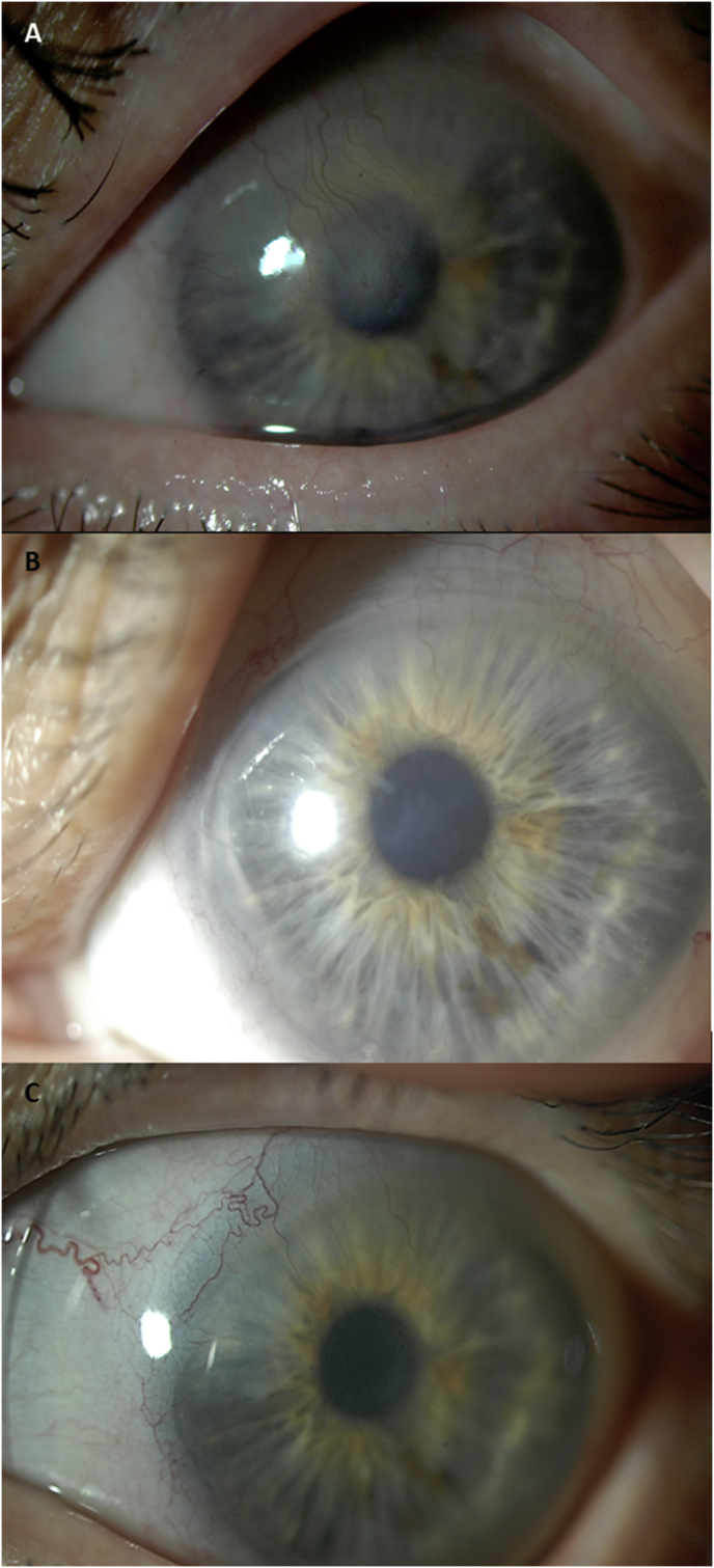
Purpose: To demonstrate clearing of chronic corneal opacities and improvement of visual acuity with the use of BostonSight prosthetic replacement of the ocular surface ecosystem (PROSE) treatment in ocular surface disease.
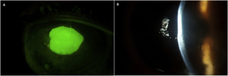
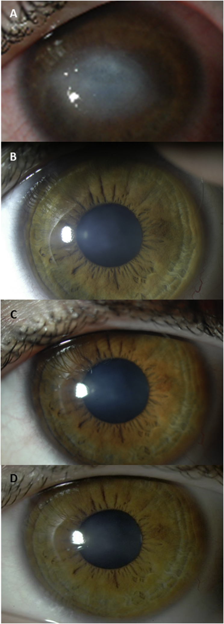
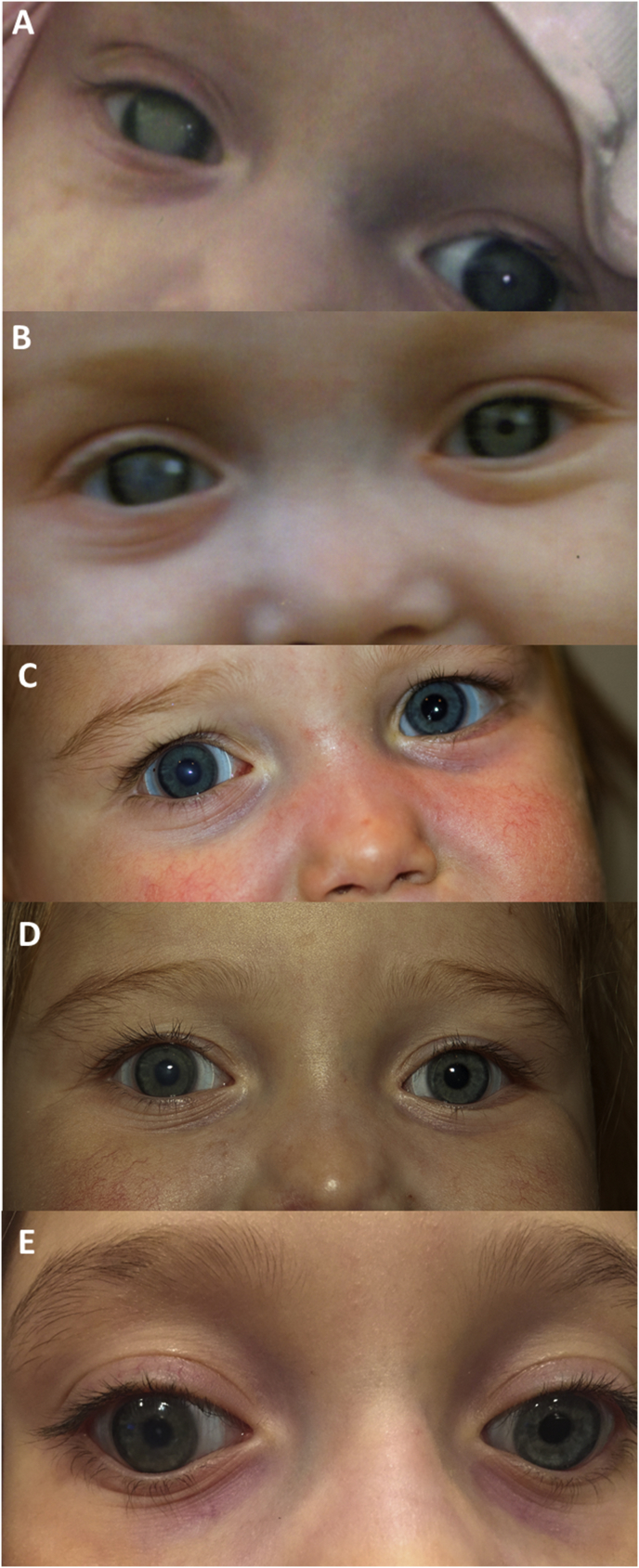
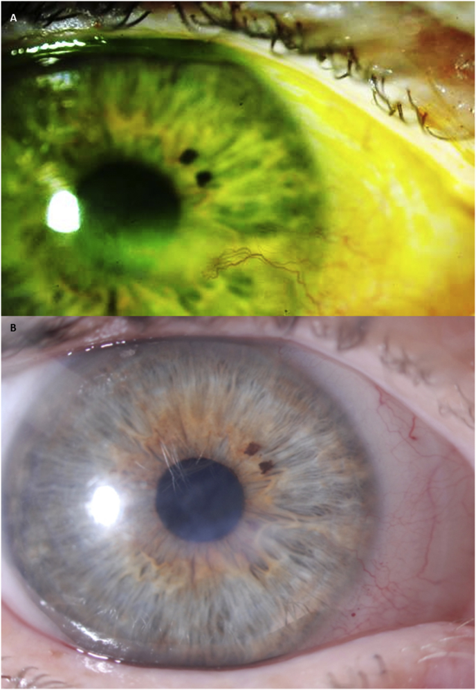
Observations: We undertook retrospective analysis of the medical records of a series of patients who underwent PROSE treatment from August 2006 to December 2014. Patients were referred for ocular surface disease of various etiologies. Primary inclusion criterion was corneal opacity that improved with PROSE treatment. Patients were excluded if topical steroids or adjuvant therapy used once PROSE treatment was initiated. Underlying disease, prior treatment, clinical presentation, and clinical course were extracted from the medical record. Four patients are included in this series. There were three females and one male; median age at time of treatment initiation was 30 years (range = 0.5-58 years). Median duration of PROSE treatment at time of retrospective analysis was 3.5 years (range = 1-8 years). Two cases had corneal opacification in the context of neurotrophic keratopathy: a unilateral case due to presumed herpes simplex keratitis and a bilateral case due to congenital corneal anesthesia associated with familial dysautonomia. One case had corneal opacity from exposure related to seventh nerve palsy, and one had corneal opacification associated with recurrent surface breakdown, neurotrophic keratopathy, and limbal stem deficiency of uncertain etiology. After consistent wear of prosthetic devices used in PROSE treatment for support of the ocular surface, visual acuity improved and clearing of the opacities was observed, without use of topical steroids or adjuvant therapy.
Conclusions and importance: These cases demonstrate clearing of chronic corneal opacity with PROSE treatment for ocular surface disease. This clearing can occur with no adjuvant therapy, suggesting that restoration of ocular surface function and integrity allows for corneal remodeling.
Keywords: Corneal scar; Dry eye syndrome; Ocular surface disease; Opacity; PROSE treatment; Scleral lenses; Scleral prosthetic devices.
Figures
Similar articles
-
Regression of corneal opacity and neovascularization in Stevens-Johnson syndrome and Toxic Epidermal Necrolysis with the use of prosthetic replacement of the ocular surface ecosystem (PROSE) treatment.Am J Ophthalmol Case Rep. 2022 Apr 14;26:101520. doi: 10.1016/j.ajoc.2022.101520. eCollection 2022 Jun. Am J Ophthalmol Case Rep. 2022. PMID: 35464679 Free PMC article.
-
Long-term outcome of using Prosthetic Replacement of Ocular Surface Ecosystem (PROSE) as a drug delivery system for bevacizumab in the treatment of corneal neovascularization.Ocul Surf. 2019 Jan;17(1):134-141. doi: 10.1016/j.jtos.2018.11.008. Epub 2018 Nov 20. Ocul Surf. 2019. PMID: 30468876 Free PMC article.
-
Assessment of the Prosthetic Replacement of Ocular Surface Ecosystem (PROSE) scleral lens on visual acuity for corneal irregularity and ocular surface disease.Ocul Surf. 2018 Apr;16(2):254-258. doi: 10.1016/j.jtos.2018.01.003. Epub 2018 Feb 6. Ocul Surf. 2018. PMID: 29425812
-
Long-Term Descemetocele Management With Prosthetic Replacement of the Ocular Surface Ecosystem (PROSE) Treatment.Eye Contact Lens. 2020 Mar;46(2):e7-e10. doi: 10.1097/ICL.0000000000000602. Eye Contact Lens. 2020. PMID: 30985491 Review.
-
Central Toxic Keratopathy After Contact Lens Wear and Mechanical Debridement: Clinical Characteristics, and Visual and Corneal Tomographic Outcomes.Eye Contact Lens. 2019 Jul;45(4):e15-e23. doi: 10.1097/ICL.0000000000000575. Eye Contact Lens. 2019. PMID: 31241605 Review.
Cited by
-
In vivo biocompatibility evaluation of in situ-forming polyethylene glycol-collagen hydrogels in corneal defects.Sci Rep. 2021 Dec 13;11(1):23913. doi: 10.1038/s41598-021-03270-3. Sci Rep. 2021. PMID: 34903788 Free PMC article.
-
Two cases of therapeutic scleral lenses for KID syndrome.Am J Ophthalmol Case Rep. 2025 Jan 19;37:102261. doi: 10.1016/j.ajoc.2025.102261. eCollection 2025 Mar. Am J Ophthalmol Case Rep. 2025. PMID: 39927073 Free PMC article.
-
Regression of corneal opacity and neovascularization in Stevens-Johnson syndrome and Toxic Epidermal Necrolysis with the use of prosthetic replacement of the ocular surface ecosystem (PROSE) treatment.Am J Ophthalmol Case Rep. 2022 Apr 14;26:101520. doi: 10.1016/j.ajoc.2022.101520. eCollection 2022 Jun. Am J Ophthalmol Case Rep. 2022. PMID: 35464679 Free PMC article.
-
Strategic combination of cultivated oral mucosal epithelial transplantation and postoperative limbal-rigid contact lens-wear for end-stage ocular surface disease: a retrospective cohort study.Br J Ophthalmol. 2024 Jul 23;108(8):1177-1183. doi: 10.1136/bjo-2023-323617. Br J Ophthalmol. 2024. PMID: 37918892 Free PMC article.
-
Familial dysautonomia.Clin Auton Res. 2023 Jun;33(3):269-280. doi: 10.1007/s10286-023-00941-1. Epub 2023 May 19. Clin Auton Res. 2023. PMID: 37204536 Review.
References
Publication types
LinkOut - more resources
Full Text Sources
Other Literature Sources
