. 2018 May 2;63(9):095010.
doi: 10.1088/1361-6560/aab79b.

The influence of medium elasticity on the prediction of histotripsy-induced bubble expansion and erythrocyte viability

Affiliations

The influence of medium elasticity on the prediction of histotripsy-induced bubble expansion and erythrocyte viability

Kenneth B Bader. Phys Med Biol. .

Abstract

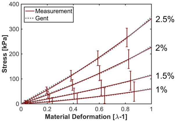
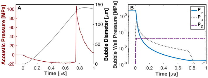
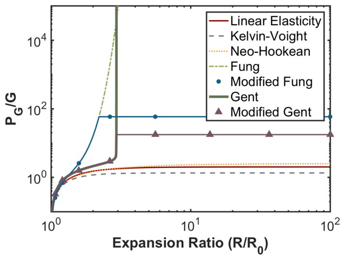
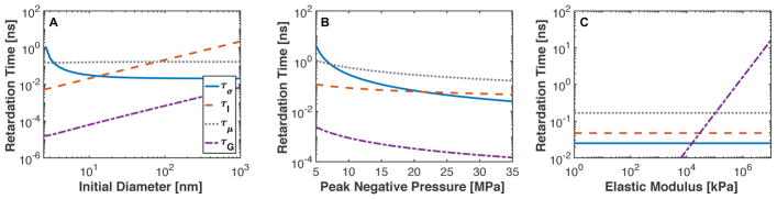
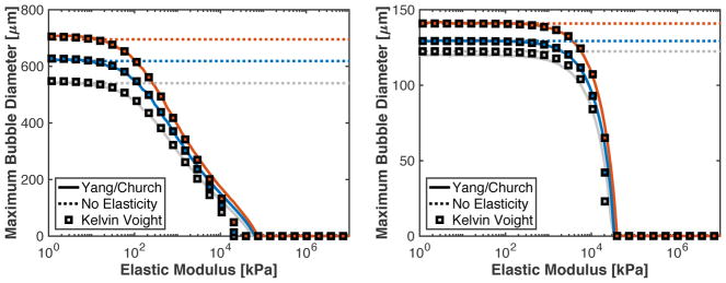
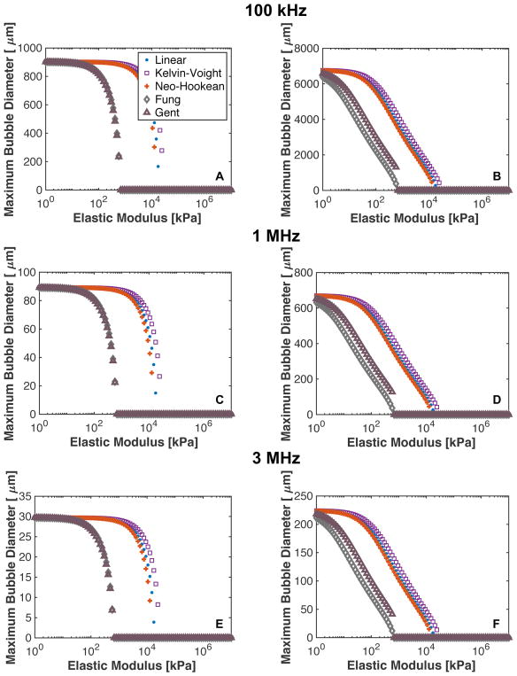
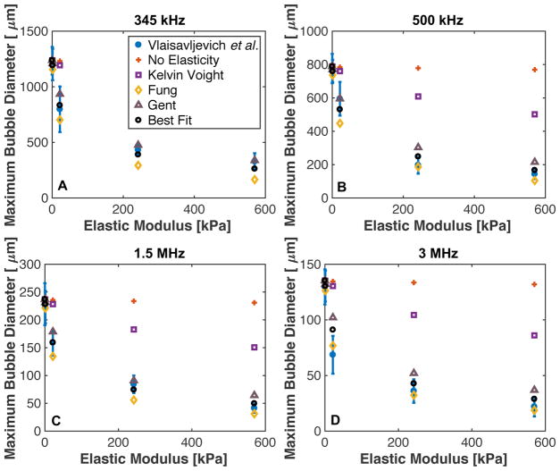
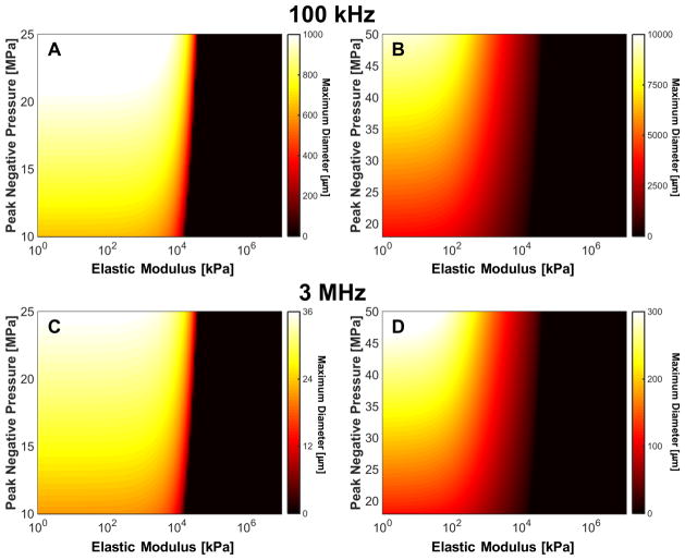
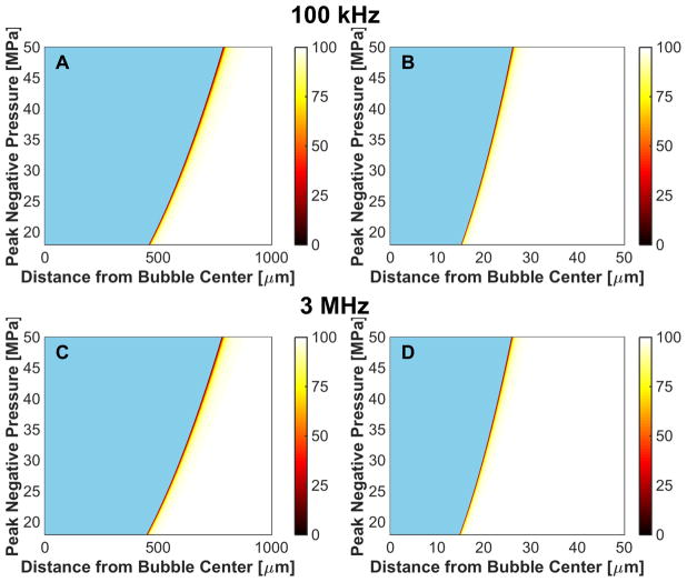
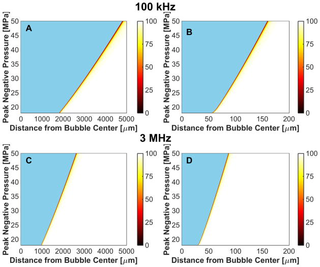
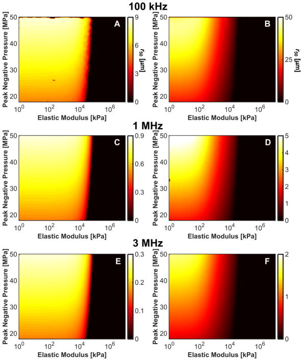
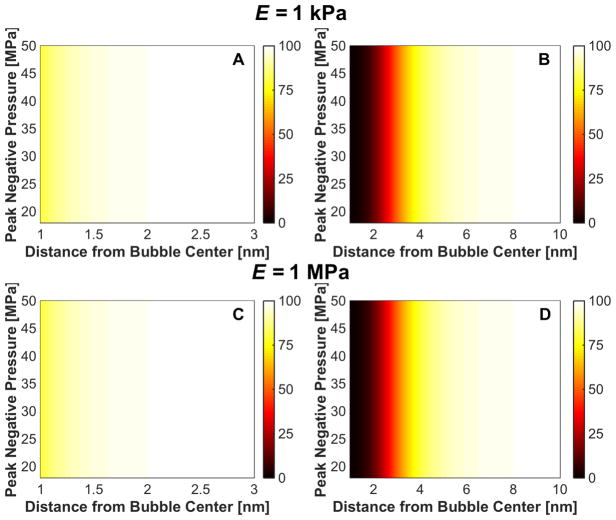
Histotripsy is a form of therapeutic ultrasound that liquefies tissue mechanically via acoustic cavitation. Bubble expansion is paramount in the efficacy of histotripsy therapy, and the cavitation dynamics are strongly influenced by the medium elasticity. In this study, an analytic model to predict histotripsy-induced bubble expansion in a fluid was extended to include the effects of medium elasticity. Good agreement was observed between the predictions of the analytic model and numerical computations utilizing highly nonlinear excitations (shock-scattering histotripsy) and purely tensile pulses (microtripsy). No bubble expansion was computed for either form of histotripsy when the elastic modulus was greater than 20 MPa and the peak negative pressure was less than 50 MPa. Strain in the medium due to the expansion of a single bubble was also tabulated. The viability of red blood cells was calculated as a function of distance from the bubble wall based on empirical data of impulsive stretching of erythrocytes. Red blood cells remained viable at distances further than 44 µm from the bubble wall. As the medium elasticity increased, the distance over which bubble expansion-induced strain influenced red blood cells was found to decrease sigmoidally. These results highlight the relationship between tissue elasticity and the efficacy of histotripsy. In addition, an upper medium elasticity limit was identified, above which histotripsy may not be effective for tissue liquefaction.


Figures

Fig. A1
Fig. A1
Measured stress versus material deformation in agar phantoms (Table 1 in (Barrangou et al 2006)) and least-squares fits of the Gent model, A1, to the measured stress to determine parameter JM. The concentration of agar is noted along the side of the figure.
Fig. 1
Fig. 1
(A) Expansion of a 20-nm cavitation nucleus to a shock-scattering histotripsy pulse. (B) Pressure components due to surface tension (Pσ), viscosity (Pμ), and elasticity (PG), corresponding to the second, third, and forth terms in (2).
Fig. 2
Fig. 2
Elastic pressure contribution as a function of the bubble expansion ratio for elastic models described in Table 1.
Fig. 3
Fig. 3
Bubble expansion delays due to surface tension (τσ), fluid inertia ( τI), viscosity (τμ), and tissue elasticity (τG) as a function of (A) initial bubble diameter with peak negative pressure (23 MPa) and elastic modulus (90 kPa), (B) peak negative pressure with fixed initial bubble diameter (20 nm) and elastic modulus (90 kPa), and (C) elastic modulus with fixed initial bubble diameter (20 nm) and peak negative pressure (23 MPa). The legend for each component of time delay is shown in panel A.
Fig. 4
Fig. 4
(A) Predicted maximum bubble size as a function of elastic modulus for microtripsy pulses with 24.9 (gray), 29.3 (blue), and 33.6 MPa (red) peak negative pressures. (B) Predicted maximum bubble size as a function of elastic modulus for a single cycle shock-scattering histotripsy pulse with peak negative/peak positive pressures of 14.5/88.9 (gray), 15.8/98.8 (blue), and 18.3/107.0 MPa (red) peak negative/peak positive pressures. For both panels, the solid line is the numerical computations with the Yang/Church model with Kelvin-Vought elasticity, the dotted line is the analytic model without elasticity incorporated, and squares are the analytic theory with Kelvin-Voight elasticity incorporated.
Fig. 5
Fig. 5
(A) Bubble expansion rate over the course of a shock-scattering histotripsy pulse based on the numerical integration of the Yang/Church model (dashed line) and the analytic prediction via (9) (crosses). The peak positive/peak negative pressures were 98.8/15.8 MPa for the brown symbols, 102.8/16.9 MPa for the yellow symbols, and 107/18.3 MPa for the blue symbols. (B) Predicted axial extent of the bubble cloud initiated with a 5-μs shock-scattering histotripsy pulse as a function of the elastic modulus of the medium. The fundamental frequency of the shock scattering pulse was 1 MHz. Kelvin-Voight elasticity was used in the calculation.
Fig. 6
Fig. 6
Predicted maximum bubble diameter for linear, Kelvin-Voight, Neo-Hookean, and Fung elasticity for a single cycle shock-scattering histotripsy excitation (left column) and microtripsy excitation (right column). The fundamental frequency of the excitation was 100 kHz for panels A and B, 1 MHz for panels C and D, and 3 MHz for panels E and F. The initial nucleus for the shock scattering histotripsy excitation was 20 nm, and the peak negative pressure was 17.4 MPa. The initial nucleus size for the microtripsy pulse was 5 nm, and the peak negative pressure was 33.8 MPa.
Fig. 7
Fig. 7
Maximum size of cavitation nuclei excited by microtripsy pulse as a function of medium elasticity at (A) 345 kHz, (B) 500 kHz, (C) 1.5 MHz, and (D) 3 MHz. Blue dots are measured values (table 2 from Vlaisavljevich et al. (2013b)). Analytic calculations were computed via (3), with constants acquired from Table 1. The “Best Fit” elastic model conditions utilized the constants ξ = ξHA − 30G/P0 and pEM=P0(1+30GP0) in the analytic calculation. The legend shown in panel A labeling the elastic models used in the analytic calculation is the same for the remaining panels.
Fig. 8
Fig. 8
Predicted maximum bubble diameter for a single cycle shock-scattering histotripsy excitation (left column) and microtripsy excitation (right column). The fundamental frequency of the insonation was 100 kHz for panels A and B, and 3 MHz for panels C and D. The initial nucleus for the shock scattering histotripsy excitation was 20 nm, and initial nucleus size for the microtripsy pulse was 5 nm. Kelvin-Voight elasticity was used in the calculation.
Fig. 9
Fig. 9
Microtripsy-induced reduction in red blood cell viability due to bubble expansion, reported in terms of percent viability in the colorbar. The fundamental frequency of the insonation was 100 kHz for the left column, and 3 MHz for the right column. The elastic modulus was 1 kPa for panels A and B, and 1 MPa for panels C and D. The initial bubble diameter was 5 nm. The regions in blue represent the extent of the bubble.
Fig. 10
Fig. 10
Shock-scattering histotripsy-induced reduction in red blood cell viability due to bubble expansion, reported in terms of percent viability in the colorbar. The fundamental frequency of the insonation was 100 kHz for the left column, and 3 MHz for the right column. The elastic modulus was 1 kPa for panels A and B, and 1 MPa for panels C and D. The initial bubble diameter was 20 nm. The regions in blue represent the extent of the bubble.
Fig. 11
Fig. 11
Distance from bubble wall to 50% reduction in red blood cell viability, r50, induced by bubble expansion as a function of elastic modulus and peak negative pressure for shock-scattering histotripsy (left column) and microtripsy (right column) excitations. (A), (B) 100-kHz fundamental frequency. (C), (D) 1-MHz fundamental frequency. (E), (F), 3-MHz fundamental frequency.
Fig. 12
Fig. 12
Change in cell viability, expressed in terms of percent in the colorbar, due to strain at minimum bubble radius for microtripsy insonation (left column) and shock-scattering histotripsy insonation (right column) for medium elastic modulus of 1 kPa (panels A and B) and 1 MPa (panels C and D). The fundamental frequency of the insonation was 1 MHz for both calculations, and the initial bubble diameter was 5 nm for the microtripsy excitation and 20 nm for the shock scattering histotripsy excitation.

References

    1. Apfel RE. Acoustic cavitation prediction. J Acoust Soc Am. 1981a;69:1624–33.
    1. Apfel RE. Methods in Experimental Physics. Vol. 19. New York: Academic Press, Inc; 1981b. Acoustic Cavitation; pp. 355–411.
    1. Apfel RE. Possibility of microcavitation from diagnostic ultrasound. IEEE Trans Ultrason Ferro Freq Control. 1986;33:139–42. - PubMed
    1. Apfel RE, Holland CK. Gauging the likelihood of cavitation from short-pulse, low-duty cycle diagnostic ultrasound. Ultrasound Med Biol. 1991;17:179–85. - PubMed
    1. Bader KB, Holland CK. Predicting the growth of nanoscale nuclei by histotripsy pulses. Phys Med Biol. 2016:2947–66. - PMC - PubMed

Publication types