. 2018 Apr 2;128(4):1641-1656.
doi: 10.1172/JCI97248. Epub 2018 Mar 19.

Sox2 haploinsufficiency primes regeneration and Wnt responsiveness in the mouse cochlea

Affiliations

Sox2 haploinsufficiency primes regeneration and Wnt responsiveness in the mouse cochlea

Patrick J Atkinson et al. J Clin Invest. .

Abstract

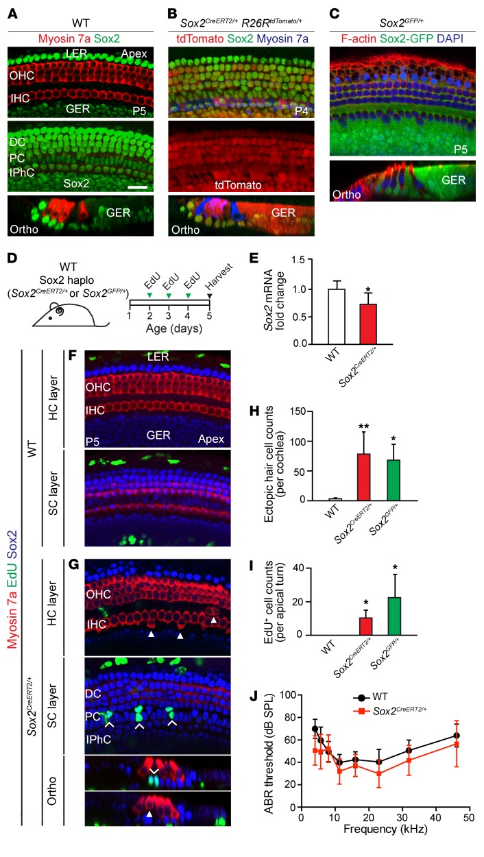
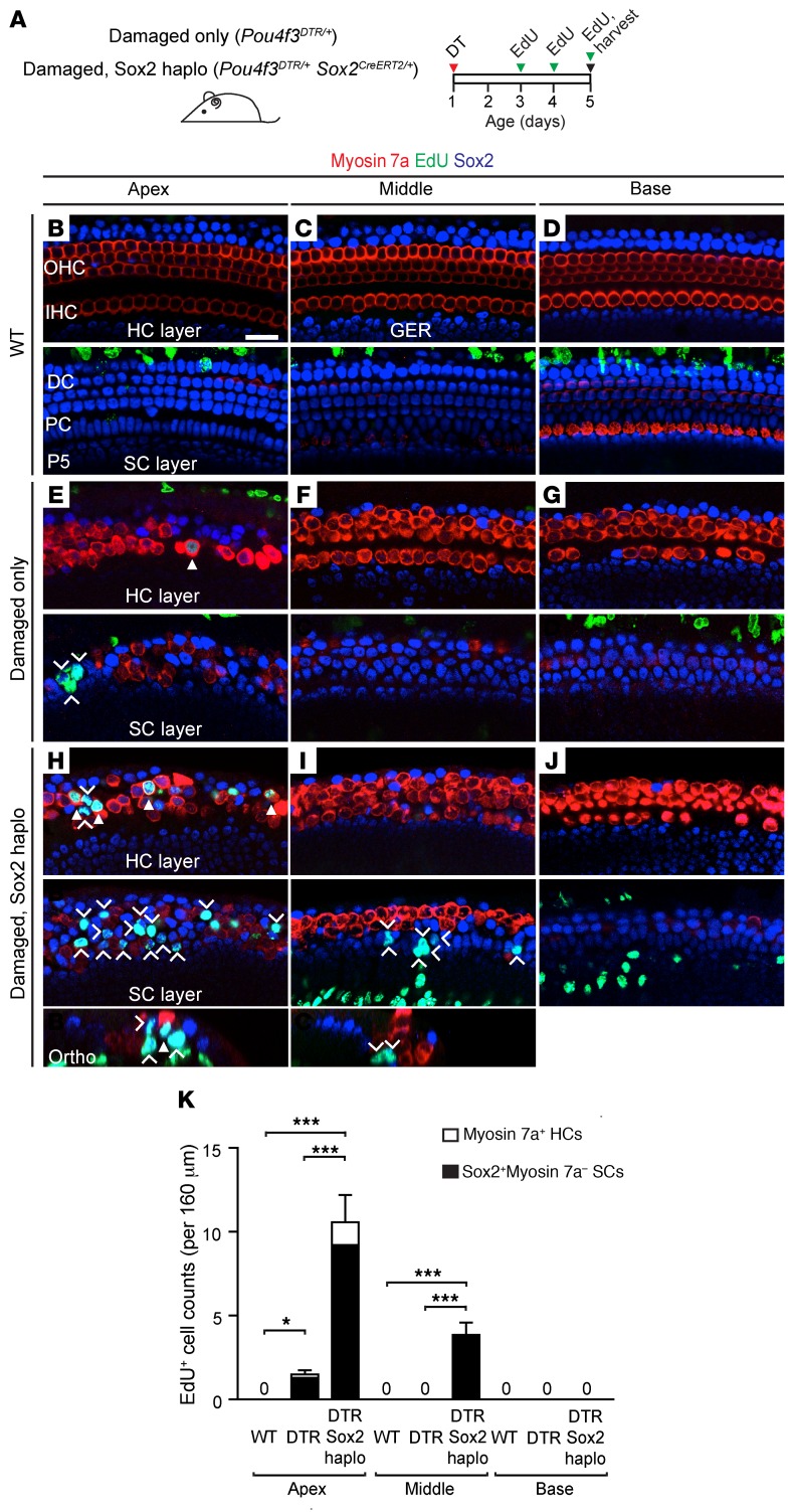
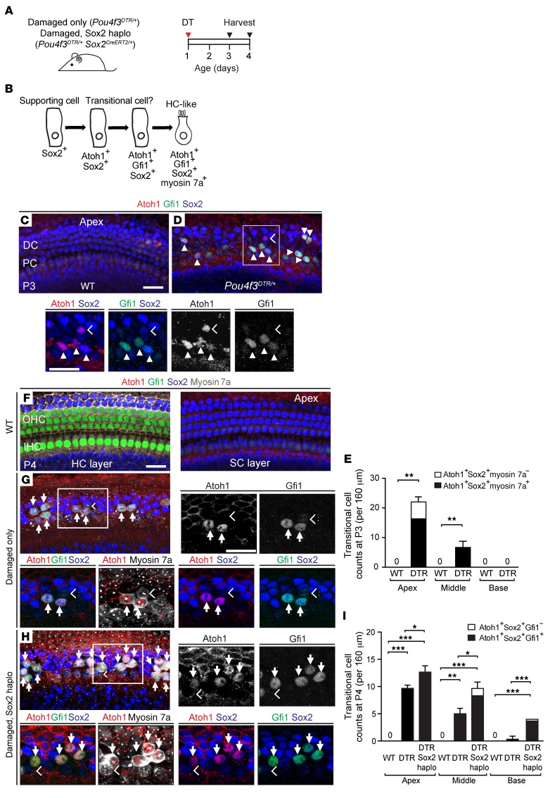
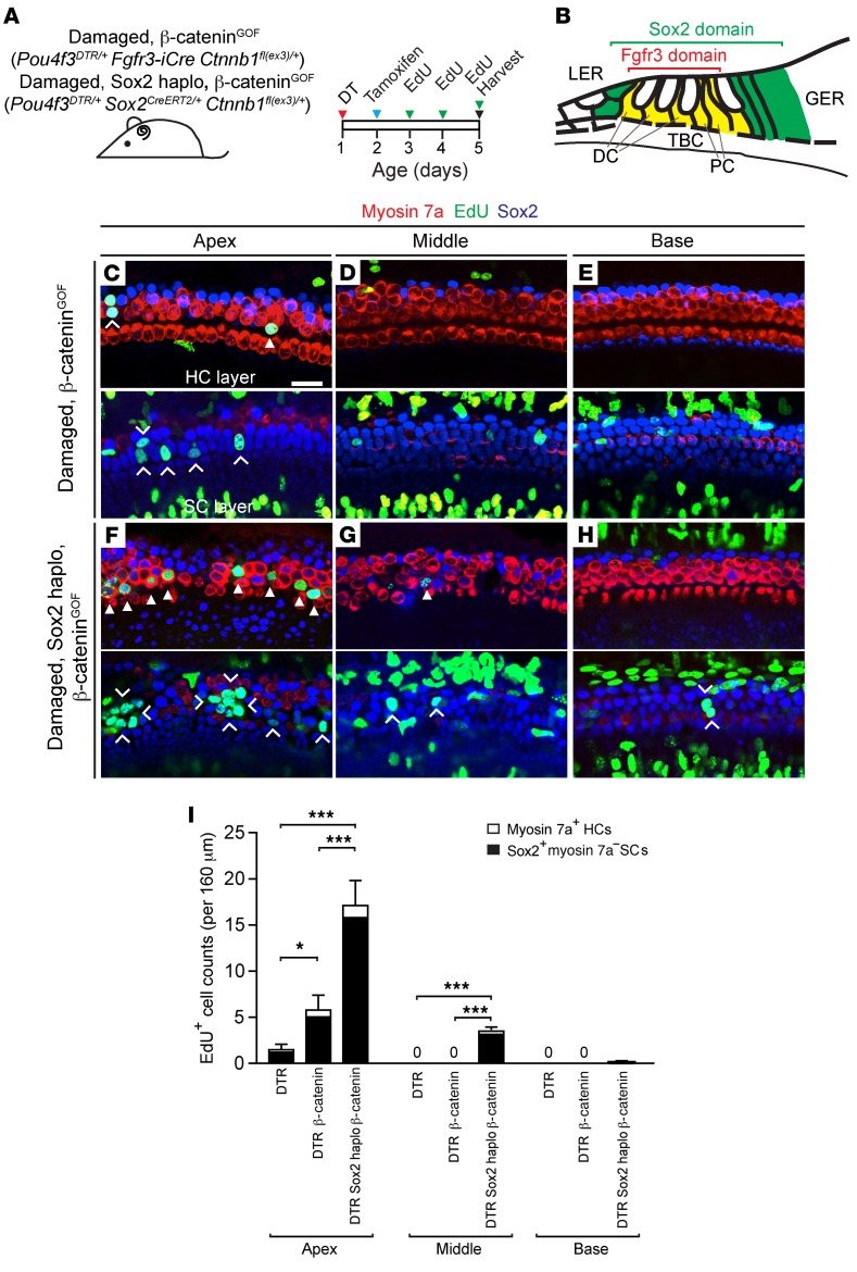
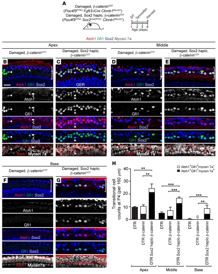
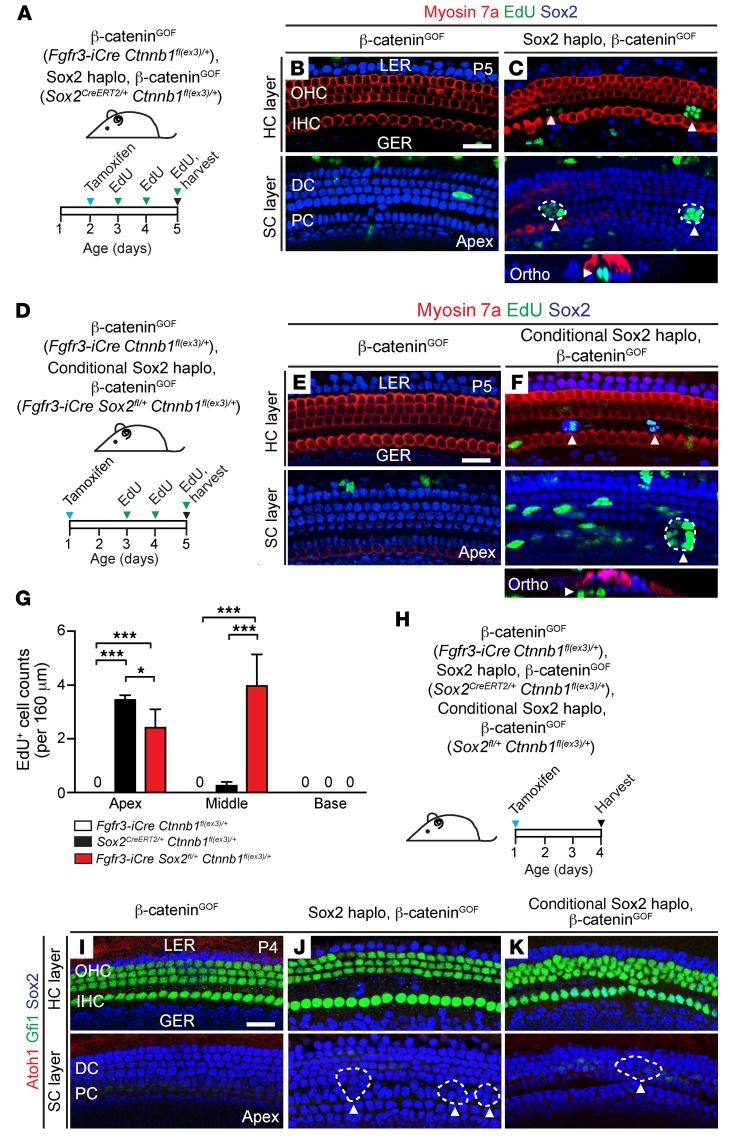
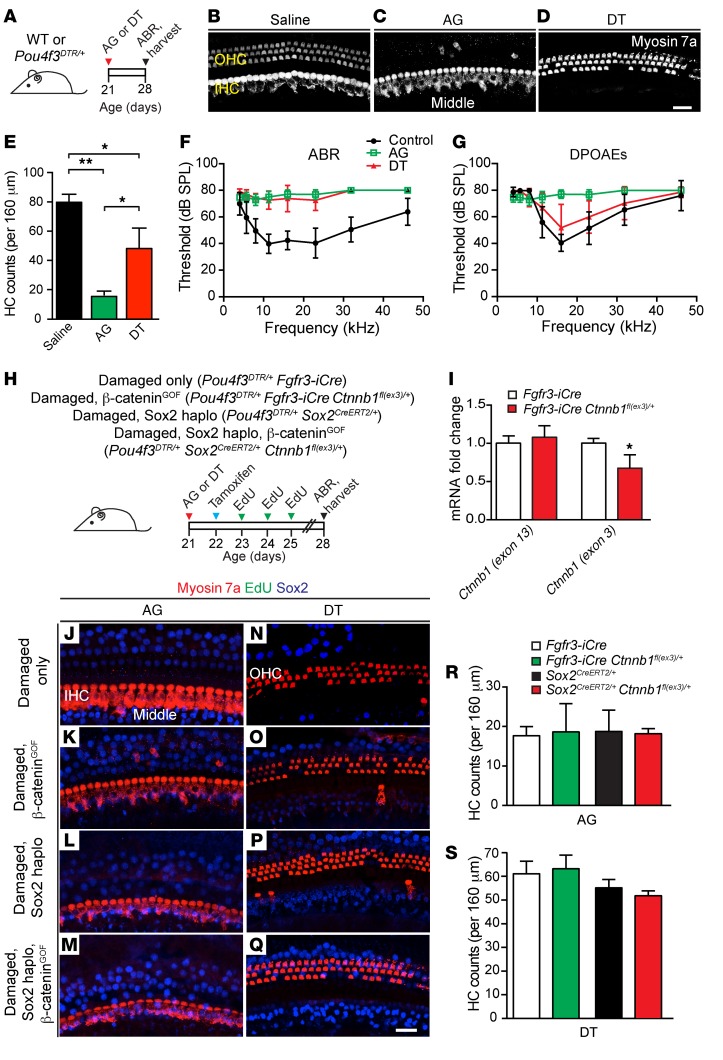
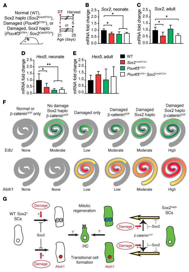
During development, Sox2 is indispensable for cell division and differentiation, yet its roles in regenerating tissues are less clear. Here, we used combinations of transgenic mouse models to reveal that Sox2 haploinsufficiency (Sox2haplo) increases rather than impairs cochlear regeneration in vivo. Sox2haplo cochleae had delayed terminal mitosis and ectopic sensory cells, yet normal auditory function. Sox2haplo amplified and expanded domains of damage-induced Atoh1+ transitional cell formation in neonatal cochlea. Wnt activation via β-catenin stabilization (β-cateninGOF) alone failed to induce proliferation or transitional cell formation. By contrast, β-cateninGOF caused proliferation when either Sox2haplo or damage was present, and transitional cell formation when both were present in neonatal, but not mature, cochlea. Mechanistically, Sox2haplo or damaged neonatal cochleae showed lower levels of Sox2 and Hes5, but not of Wnt target genes. Together, our study unveils an interplay between Sox2 and damage in directing tissue regeneration and Wnt responsiveness and thus provides a foundation for potential combinatorial therapies aimed at stimulating mammalian cochlear regeneration to reverse hearing loss in humans.

Keywords: Cell Biology; Mouse models; Mouse stem cells; Neurodegeneration; Neuroscience.


Conflict of interest statement

Conflict of interest: The authors have declared that no conflict of interest exists.

Figures

Figure 1
Figure 1. Sox2 haploinsufficiency results in continued proliferation and formation of supernumerary hair cells in the neonatal cochlea.
(A) Immunostaining of P5 WT cochlea shows Sox2 expression in Hensen’s cells, Deiters’ cells, pillar cells, and the lateral portion of the greater epithelial ridge. (B) Whole-mount preparation of cochlea from P4 Sox2CreERT2/+ R26RtdTomato/+ mice given tamoxifen on P2, showing tdTomato expression in supporting cells and some hair cells. (C) GFP+ supporting cells in the P5 Sox2GFP/+ cochlea. (D) Schematic of EdU administration to Sox2CreERT2/+ mice, Sox2GFP/+ mice, and WT littermates (once daily, P2–P4). haplo, haploinsufficient. (E) qPCR showed a significant reduction of Sox2 expression in Sox2CreERT2/+ cochleae compared with expression in WT littermates. (F) Confocal images show no EdU+ hair cells or supporting cells in the P5 WT cochlea. EdU labeling was seen in cells in the lesser epithelial ridge and greater epithelial ridge. (G) Sox2CreERT2/+ cochlea contained occasional extranumerary hair cells adjacent to inner hair cells (arrowheads). Extranumerary hair cells were noted in all cochlear turns of Sox2CreERT2/+ mice. Image shows EdU+ supporting cells (chevrons) in the apical turn. No EdU+ hair cells were noted. (H) Quantification of extranumerary hair cells in WT, Sox2CreERT2/+, and Sox2GFP/+ cochleae. (I) Quantification of EdU+ cells in WT, Sox2CreERT2/+, and Sox2GFP/+ cochleae. (J) P28 Sox2CreERT2/+ mice had normal ABR thresholds, comparable to those of their WT littermates. DC, Deiters’ cell; GER, greater epithelial ridge; HC, hair cell; IHC, inner hair cell; IP, inner pillar cell; IPhC, inner phalangeal cell; LER, lesser epithelial ridge; OHC, outer hair cell; OP, outer pillar cell; Ortho, orthogonal view; PC, pillar cell; SC, supporting cell. Data represent the mean ± SD. *P < 0.05 and **P < 0.01, by 2-tailed Student’s t test. n = 3–8. Scale bar: 20 μm.
Figure 2
Figure 2. Reduced Sox2 levels enhance and expand the domain of proliferation in the damaged neonatal mouse cochlea.
(A) Schematic of hair cell ablation in neonatal cochlea. Briefly, Pou4f3DTR/+ and Pou4f3DTR/+ Sox2CreERT2/+ mice were injected with DT on P1 to induce hair cell loss, followed by administration of EdU (P3–P5), and cochleae were examined on P5. (BD) No EdU+ hair cells or supporting cells were found in any of the 3 WT cochlear turns. (EG) In the Pou4f3DTR/+ cochlea, after DT-induced hair cell damage, EdU+myosin 7a+ hair cells (arrowhead) and some EdU+Sox2+ supporting cells (chevrons) were observed in the apical turn, but not in the basal or middle turns. (HJ) In Pou4f3DTR/+ Sox2CreERT2/+ cochlea, there was robust EdU labeling of both myosin 7a+ hair cells (arrowheads) and Sox2+ supporting cells (chevrons) in the apical turn. EdU+Sox2+ supporting cells were also found in the middle turn. (K) Quantification of EdU+myosin 7a+ hair cells and myosin 7aSox2+ supporting cells per cochlear turn. Data represent the mean ± SD. *P < 0.05 and ***P < 0.001, by 1-way ANOVA with Holm-Sidak multiple comparisons test. n = 5–6. Scale bar: 20 μm.
Figure 3
Figure 3. Sox2 reduction enhances transitional cell formation in the damaged neonatal mouse cochlea.
(A) Schematic of hair cell ablation. P1 Pou4f3DTR/+ and Pou4f3DTR/+ Sox2CreERT2/+ pups were injected with DT, and cochleae were examined on P3 and P4. (B) Cartoon depicts supporting cells forming transitional cells during regeneration. (C) Confocal images of P3 WT cochlea show no Atoh1 or Gfi1 expression in supporting cells. (D) After hair cell damage in the Pou4f3DTR/+ cochlea, some Sox2+ supporting cells expressed Atoh1 (chevrons) and Gfi1 (arrowheads), both early hair cell markers. All Gfi1+Sox2+ supporting cells expressed Atoh1, but some Atoh1+Sox2+ supporting cells did not express Gfi1. (E) Quantification of transitional cells (Atoh1+Sox2+ and Atoh1+Sox2+Gfi1+) from WT and Pou4f3DTR/+ cochleae. (F) P4 WT cochlea showed Gfi1 expression limited to hair cells and no Atoh1 or Gfi1 expression in Sox2+ supporting cells. Atoh1 was absent in hair cells. (G) After DT-induced hair cell loss in Pou4f3DTR/+ cochlea, Gfi1 was downregulated in the remaining myosin 7a+ hair cells. Many transitional cells (arrows) (Atoh1+Sox2+myosin 7a+Gfi1+) were detected in all 3 cochlear turns. Like the P3 Pou4f3DTR/+ cochlea, all transitional cells expressed Atoh1 and Sox2. In contrast to the P3 Pou4f3DTR/+ cochlea, most transitional cells expressed myosin 7a by P4. Myosin7a+ cells with no expression of Sox2, Atoh1, or Gfi1 (chevron) likely represent surviving hair cells (G and H). (H) In the P4 Pou4f3DTR/+ Sox2CreERT2/+ cochlea, there were noticeably more transitional cells (arrows). (I) Quantification of transitional cells (Atoh1+Sox2+myosin 7a+Gfi1+ and Atoh1+Sox2+Gfi1+) in WT, Pou4f3DTR/+, and Pou4f3DTR/+ Sox2CreERT2/+ cochleae. Hair cell ablation led to a significantly greater number of transitional cells in each cochlear turn compared with that seen in controls. There were significantly more transitional cells detected in each turn of damaged, Sox2haplo cochleae than in the damaged-only cochleae. The SD of transitional cell counts in the basal turn of damaged, Sox2haplo cochleae is zero. Data represent the mean ± SD. *P < 0.05, **P < 0.01, and ***P < 0.001, by 1-way ANOVA with Holm-Sidak multiple comparisons test. n = 3. Scale bars: 20 μm.
Figure 4
Figure 4. β-Catenin stabilization and Sox2 haploinsufficiency coordinate to increase mitotic regeneration in the damaged neonatal mouse cochlea.
(A) Pou4f3DTR/+ Fgfr3-iCre Ctnnb1fl(ex3)/+ and Pou4f3DTR/+ Sox2CreERT2/+ Ctnnb1fl(ex3)/+ pups were injected with DT on P1, tamoxifen on P2, and EdU daily (P3–P5), and cochleae were collected on P5. (B) Schematic depicting the domains of Fgfr3 and Sox2 expression in the neonatal mouse cochlea. (CE) Confocal images of cochleae from P5 Pou4f3DTR/+ Fgfr3-iCre Ctnnb1fl(ex3)/+ mice showing EdU+myosin 7a+ hair cells (arrowhead) and EdU+Sox2+ supporting cells (chevrons) in the apical turn, but not in the middle or basal turn. Note that many EdU+Sox2 cells resided outside the sensory epithelium. (FH) In Pou4f3DTR/+ Sox2CreERT2/+ Ctnnb1fl(ex3)/+ cochlea, there was a robust increase in the number of EdU+myosin 7a+ hair cells (arrowheads) and Sox2+ supporting cells (chevrons) in the apical turn. As with Pou4f3DTR/+ Sox2CreERT2/+ cochlea, EdU+ supporting cells were noted in the middle turns and occasionally in the basal turns. Many EdU+Sox2 cells outside the sensory epithelium were also noted. (I) Quantification of EdU+myosin 7a+ hair cells and EdU+Sox2+ supporting cells in Pou4f3DTR/+, Pou4f3DTR/+ Fgfr3-iCre Ctnnb1fl(ex3)/+, and Pou4f3DTR/+ Sox2CreERT2/+ Ctnnb1fl(ex3)/+ cochleae. Data represent the mean ± SD. *P < 0.05 and ***P < 0.001, by 1-way ANOVA with Holm-Sidak multiple comparisons test. n = 3–5. Scale bar: 20 μm.
Figure 5
Figure 5. β-Catenin stabilization and Sox2 haploinsufficiency coordinate to increase transitional cell formation in the damaged neonatal mouse cochlea.
(A) Pou4f3DTR/+ Fgfr3-iCre Ctnnb1fl(ex3)/+ and Pou4f3DTR/+ Sox2CreERT2/+ Ctnnb1fl(ex3)/+ mice were injected with DT on P1, followed by tamoxifen administration on P2, and cochleae were examined on P4. (B, D, and F) As with Pou4f3DTR/+ cochlea, some transitional cells (Atoh1+Gfi1+Sox2+myosin 7a+, arrows) were detected in the apical and middle turns of the Pou4f3DTR/+ Fgfr3-iCre Ctnnb1fl(ex3)/+ cochlea. Some Atoh1+Gfi1+Sox2+ (myosin 7a) cells (arrowheads), which were rarely seen in the Pou4f3DTR/+ cochlea, were also noted in the supporting cell layer of apical and middle turns. Some myosin 7a+Atoh1Gfi1 hair cells (chevrons) were also observed in all 3 turns and were presumed to be surviving hair cells. (C, E, and G) In the Pou4f3DTR/+ Sox2CreERT2/+ Ctnnb1fl(ex3)/+ cochlea, a marked increase in transitional cells was observed in all 3 turns. (H) Quantification revealed that significantly more transitional cells were detected in all 3 turns in the Pou4f3DTR/+ Sox2CreERT2/+ Ctnnb1fl(ex3)/+ cochleae than in either Pou4f3DTR/+ or Pou4f3DTR/+ Fgfr3-iCre Ctnnb1fl(ex3)/+ cochleae. Data represent the mean ± SD. **P < 0.01 and ***P < 0.001, by 1-way ANOVA with Holm-Sidak multiple comparisons test. n = 3. Scale bar: 20 μm.
Figure 6
Figure 6. Sox2 haploinsufficiency acts as a permissive signal for β-catenin–induced proliferation in the undamaged neonatal cochlea.
(A) Schematic of the experimental paradigm. Fgfr3-iCre Ctnnb1fl(ex3)/+ and Sox2CreERT2/+ Ctnnb1fl(ex3)/+ pups were given tamoxifen on P2, followed by daily administration of EdU (P3–P5), and cochleae were examined on P5. (B) No EdU+ cells were detected in the organ of Corti in cochleae from Fgfr3-iCre Ctnnb1fl(ex3)/+ mice treated with tamoxifen on P2. (C) EdU+ supporting cells but not hair cells arranged as foci (dashed lines, arrowhead) were found in P5 Sox2CreERT2/+ Ctnnb1fl(ex3)/+ cochleae. (D) Fgfr3-iCre Ctnnb1fl(ex3)/+ and Fgfr3-iCre Sox2fl/+ Ctnnb1fl(ex3)/+ pups were given tamoxifen on P1, followed by EdU administration daily (P3–P5), and cochleae were examined on P5. (E) The apical turn of Fgfr3-iCre Ctnnb1fl(ex3)/+ cochleae revealed no EdU+ hair cells or supporting cells. (F) Like Sox2CreERT2/+ Ctnnb1fl(ex3)/+ cochlea, but in contrast to Fgfr3-iCre Ctnnb1fl(ex3)/+ cochlea, EdU+ cells arranged as clusters (dashed lines, arrowheads) were found in Fgfr3-iCre Ctnnb1fl(ex3)/+ Sox2fl/+ cochlea (tamoxifen was given on P1). (G) Quantification of EdU+ cells. The differences in EdU+ cells between Sox2haplo and conditional Sox2haplo models can be attributed to the timing or degrees of Sox2 partial deletion. (H) Fgfr3-iCre Ctnnb1fl(ex3)/+, Sox2CreERT2/+ Ctnnb1fl(ex3)/+, and Fgfr3-iCre Sox2fl/+ Ctnnb1fl(ex3)/+ pups were injected with tamoxifen on P1, and cochleae were harvested on P4. (I) Gfi1-labeled hair cells were present, but no Atoh1- or Gfi1-labeled Sox2+ supporting cells were detected in Fgfr3-iCre Ctnnb1fl(ex3)/+ cochlea. (J) No Atoh1- or Gfi1-labeled Sox2+ supporting cells were detected in Sox2CreERT2/+ Ctnnb1fl(ex3)/+ cochlea, although foci-like clusters were still noted in the pillar cell region (dashed lines, arrowheads). (K) Neither Atoh1- nor Gfi1-labeled transitional cells were detected in Fgfr3-iCre Ctnnb1fl(ex3)/+ Sox2fl/+ cochlea (tamoxifen was administered on P1), although foci-like clusters could still be observed (dashed line, arrowhead). Data represent the mean ± SD. *P < 0.05 and ***P < 0.001, by 1-way ANOVA with Holm-Sidak multiple comparisons test. n = 3. Scale bars: 20 μm.
Figure 7
Figure 7. Sox2 haploinsufficiency and β-catenin stabilization do not induce mitotic hair cell regeneration in the damaged adult cochlea.
(A) Schematic showing the use of AG (sisomicin combined with furosemide) or DT to damage the mature cochlea in WT and Pou4f3DTR/+ mice. (BD) Saline-treated mice containing a full complement of myosin 7a+ cochlear inner hair cells and outer hair cells. A loss of outer hair cells after AG treatment and of inner hair cells after DT treatment was observed. Some outer hair cell loss after DT treatment was observed. (E) Quantification revealed a significant decrease in hair cell numbers in both damage paradigms. (F and G) ABR thresholds were significantly higher in both the AG- and DT-treated animals as compared with controls. A significant threshold shift was also observed in the DPOAEs in AG-treated animals, but not in the animals treated with DT. (H) Schematic of transgenic mouse models and experimental timeline. Cochleae were damaged on P21, tamoxifen was given on P22, followed by EdU administration from P23 to P25, and the animals were sacrificed after ABR on P28. (I) qPCR showing a significant reduction of Ctnnb1 (exon3) mRNA expression but not of Ctnnb1 (exon 13) mRNA expression in cochleae from Fgfr3-iCre Ctnnb1fl(ex3)/+ mice. (JM) No EdU+Sox2+ supporting cells or myosin 7a+ hair cells were detected after AG treatment in any of the genotypes examined. A persistent loss of outer hair cells was seen in these cochleae, without any new Sox2+myosin 7a+ hair cells. (NQ) There were no EdU+ hair cells or supporting cells after DT treatment or formation of new Sox2+myosin 7a+ hair cells in any of the mouse cohorts. (R and S) Quantification of hair cells in AG- and DT-treated cochleae showing no change in hair cell numbers among genotypes. Data represent the mean ± SD. *P < 0.05 and **P < 0.01, by 2-tailed Student’s t test or 1-way ANOVA with Holm-Sidak multiple comparisons test. n = 3–11. Scale bars: 20 μm.
Figure 8
Figure 8. qPCR of Notch and Wnt target genes in damaged and Sox2-haploinsufficient cochleae.
(A) Neonatal (P1) and mature (P21) WT, Sox2CreERT2/+, Pou4f3DTR/+, and Pou4f3DTR/+ Sox2CreERT2/+ mice were treated with DT, and cochleae were collected 4 days later. (B) A significant decrease was detected in Sox2 expression levels in the neonatal Sox2CreERT2/+, Pou4f3DTR/+, and Pou4f3DTR/+ Sox2CreERT2/+ cochleae relative to levels in WT controls. (C) Only the mature Sox2CreERT2/+ and Pou4f3DTR/+ Sox2CreERT2/+ cochleae had lower Sox2 levels than those detected in control cochleae. (D) A significant decrease in the levels of the Notch target gene Hes5 was detected in Sox2CreERT2/+, Pou4f3DTR/+, and Pou4f3DTR/+ Sox2CreERT2/+ cochleae relative to levels in WT cochleae on P5. (E) When measured on P25, no significant changes in the expression levels of Hes5 were seen relative to WT cochleae. Expression levels of Sox2 and Hes5 in the mature WT cochleae were lower than levels in P5 WT cochleae. (F) Schematics depicting the extent and patterns of proliferation and Atoh1+ transitional cells under various defined conditions. Darker colors represent more robust proliferation or the formation of transitional cells. (G) Proposed model of Sox2 and damage coordination in regulating mitotic regeneration, transitional cell formation, and Wnt responsiveness. *P < 0.05 and **P < 0.01, by 1-way ANOVA with Holm-Sidak multiple comparisons test. n = 4.

Similar articles

Cited by

References

    1. Avilion AA, Nicolis SK, Pevny LH, Perez L, Vivian N, Lovell-Badge R. Multipotent cell lineages in early mouse development depend on SOX2 function. Genes Dev. 2003;17(1):126–140. doi: 10.1101/gad.224503. - DOI - PMC - PubMed
    1. Masui S, et al. Pluripotency governed by Sox2 via regulation of Oct3/4 expression in mouse embryonic stem cells. Nat Cell Biol. 2007;9(6):625–635. doi: 10.1038/ncb1589. - DOI - PubMed
    1. Takahashi K, Yamanaka S. Induction of pluripotent stem cells from mouse embryonic and adult fibroblast cultures by defined factors. Cell. 2006;126(4):663–676. doi: 10.1016/j.cell.2006.07.024. - DOI - PubMed
    1. Yu J, et al. Induced pluripotent stem cell lines derived from human somatic cells. Science. 2007;318(5858):1917–1920. doi: 10.1126/science.1151526. - DOI - PubMed
    1. Bylund M, Andersson E, Novitch BG, Muhr J. Vertebrate neurogenesis is counteracted by Sox1-3 activity. Nat Neurosci. 2003;6(11):1162–1168. doi: 10.1038/nn1131. - DOI - PubMed

Publication types

MeSH terms