

A Mouse Model of Intestinal Partial Obstruction
- PMID: 29553517
- PMCID: PMC5931449
- DOI: 10.3791/57381
A Mouse Model of Intestinal Partial Obstruction
Abstract
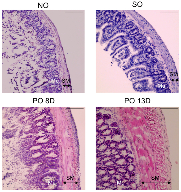
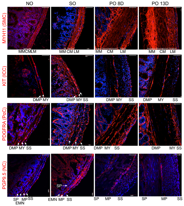
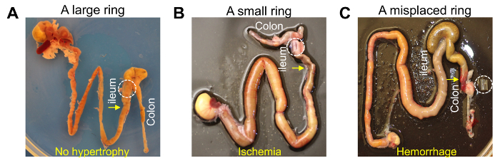
Intestinal obstructions, that impede or block peristaltic movement, can be caused by abdominal adhesions and most gastrointestinal (GI) diseases including tumorous growths. However, the cellular remodeling mechanisms involved in, and caused by, intestinal obstructions are poorly understood. Several animal models of intestinal obstructions have been developed, but the mouse model is the most cost/time effective. The mouse model uses the surgical implantation of an intestinal partial obstruction (PO) that has a high mortality rate if it is not performed correctly. In addition, mice receiving PO surgery fail to develop hypertrophy if an appropriate blockade is not used or not properly placed. Here, we describe a detailed protocol for PO surgery which produces reliable and reproducible intestinal obstructions with a very low mortality rate. This protocol utilizes a surgically placed silicone ring that surrounds the ileum which partially blocks digestive movement in the small intestine. The partial blockage makes the intestine become dilated due to the halt of digestive movement. The dilation of the intestine induces smooth muscle hypertrophy on the oral side of the ring that progressively develops over 2 weeks until it causes death. The surgical PO mouse model offers an in vivo model of hypertrophic intestinal tissue useful for studying pathological changes of intestinal cells including smooth muscle cells (SMC), interstitial cells of Cajal (ICC), PDGFRα+, and neuronal cells during the development of intestinal obstruction.


References
-
- Millat B, Guillon F. Physiopathology and principles of intensive care in intestinal obstructions. Rev Prat. 1993;43:667–672. - PubMed
-
- Tonelli P. New developments in Crohn's disease: solution of doctrinal mysteries and reinstatement as a surgically treatable disease. 1. The process is not a form of enteritis but lymphedema contaminated by intestinal contents. Chir Ital. 2000;52:109–121. - PubMed
-
- Dvorak D, Adamova Z, Bar T, Slovacek R. Internal hernia as a cause of small bowel obstruction. Rozhl Chir. 2017;96:34–36. - PubMed
Publication types
MeSH terms
Grants and funding
LinkOut - more resources
Full Text Sources
Other Literature Sources
Medical
