

Response Changes During Insertion of a Cochlear Implant Using Extracochlear Electrocochleography
- PMID: 29554036
- PMCID: PMC6139286
- DOI: 10.1097/AUD.0000000000000571
Response Changes During Insertion of a Cochlear Implant Using Extracochlear Electrocochleography
Abstract
Objectives: Electrocochleography is increasingly being utilized as an intraoperative monitor of cochlear function during cochlear implantation (CI). Intracochlear recordings from the advancing electrode can be obtained through the device by on-board capabilities. However, such recordings may not be ideal as a monitor because the recording electrode moves in relation to the neural and hair cell generators producing the responses. The purposes of this study were to compare two extracochlear recording locations in terms of signal strength and feasibility as intraoperative monitoring sites and to characterize changes in cochlear physiology during CI insertion.
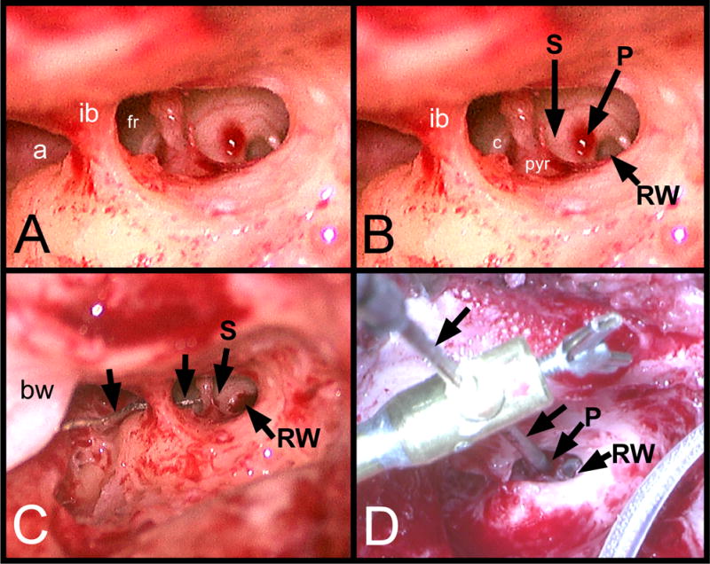
Design: In 83 human subjects, responses to 90 dB nHL tone bursts were recorded both at the round window (RW) and then at an extracochlear position-either adjacent to the stapes or on the promontory just superior to the RW. Recording from the fixed, extracochlear position continued during insertion of the CI in 63 cases.
Results: Before CI insertion, responses to low-frequency tones at the RW were roughly 6 dB larger than when recording at either extracochlear site, but the two extracochlear sites did not differ from one another. During CI insertion, response losses from the promontory or adjacent to the stapes stayed within 5 dB in ≈61% (38/63) of cases, presumably indicating atraumatic insertions. Among responses which dropped more than 5 dB at any time during CI insertion, 12 subjects showed no response recovery, while in 13, the drop was followed by partial or complete response recovery by the end of CI insertion. In cases with recovery, the drop in response occurred relatively early (<15 mm insertion) compared to those where there was no recovery. Changes in response phase during the insertion occurred in some cases; these may indicate a change in the distributions of generators contributing to the response.
Conclusions: Monitoring the electrocochleography during CI insertion from an extracochlear site reveals insertions that are potentially atraumatic, show interaction with cochlear structures followed by response recovery, or show interactions such that response losses persist to the end of recording.
Conflict of interest statement
Conflicts of Interest:
CG, TF, SP, KB, and HP declare that their involvement in research was conducted in the absence of any commercial or financial relationships that could be construed as a potential conflict of interest. CB is a consultant for Advanced Bionics and Cochlear Corporation, and OA is a consultant for MED-EL and Advanced Bionics. OA and CB have equity stakes in Advanced Cochlear Diagnostics.
Figures


Similar articles
-
Intracochlear Electrocochleography: Response Patterns During Cochlear Implantation and Hearing Preservation.Ear Hear. 2019 Jul/Aug;40(4):833-848. doi: 10.1097/AUD.0000000000000659. Ear Hear. 2019. PMID: 30335669 Free PMC article.
-
Simultaneous Intra- and Extracochlear Electrocochleography During Electrode Insertion.Ear Hear. 2021 Mar/Apr;42(2):414-424. doi: 10.1097/AUD.0000000000000935. Ear Hear. 2021. PMID: 32826509
-
Intracochlear electrocochleography during cochlear implantation.Otol Neurotol. 2014 Sep;35(8):1451-7. doi: 10.1097/MAO.0000000000000451. Otol Neurotol. 2014. PMID: 24892369
-
[Application of extra- and intracochlear electrocochleography during and after cochlear implantation].HNO. 2025 Jan;73(1):14-21. doi: 10.1007/s00106-024-01481-4. Epub 2024 May 18. HNO. 2025. PMID: 38761228 Free PMC article. Review. German.
-
Correlation Between Electrocochleographic Changes During Surgery and Hearing Outcome in Cochlear Implant Recipients: A Case Report and Systematic Review of the Literature.Otol Neurotol. 2020 Mar;41(3):318-326. doi: 10.1097/MAO.0000000000002506. Otol Neurotol. 2020. PMID: 31834213
Cited by
-
Light sheet microscopy of the gerbil cochlea.J Comp Neurol. 2021 Mar;529(4):757-785. doi: 10.1002/cne.24977. Epub 2020 Aug 3. J Comp Neurol. 2021. PMID: 32632959 Free PMC article.
-
Cochlear implantation in an animal model documents cochlear damage at the tip of the implant.Braz J Otorhinolaryngol. 2022 Jul-Aug;88(4):546-555. doi: 10.1016/j.bjorl.2020.07.017. Epub 2020 Sep 20. Braz J Otorhinolaryngol. 2022. PMID: 33039317 Free PMC article.
-
Development and Characterization of an Electrocochleography-Guided Robotics-Assisted Cochlear Implant Array Insertion System.Otolaryngol Head Neck Surg. 2022 Aug;167(2):334-340. doi: 10.1177/01945998211049210. Epub 2021 Oct 5. Otolaryngol Head Neck Surg. 2022. PMID: 34609909 Free PMC article.
-
ZH-ECochG Bode Plot: A Novel Approach to Visualize Electrocochleographic Data in Cochlear Implant Users.J Clin Med. 2024 Jun 14;13(12):3470. doi: 10.3390/jcm13123470. J Clin Med. 2024. PMID: 38929998 Free PMC article.
-
Electrocochleography in Cochlear Implant Users with Residual Acoustic Hearing: A Systematic Review.Int J Environ Res Public Health. 2020 Sep 26;17(19):7043. doi: 10.3390/ijerph17197043. Int J Environ Res Public Health. 2020. PMID: 32993065 Free PMC article.
References
-
- Acharya AN, Tavora-Vieira D, Rajan GP. Using the Implant Electrode Array to Conduct Real-time Intraoperative Hearing Monitoring During Pediatric Cochlear Implantation: Preliminary Experiences. Otology & Neurotology. 2016;37(2):e148–e153. - PubMed
-
- Adunka O, Kiefer J. Impact of electrode insertion depth on intracochlear trauma. Otolaryngology-Head and Neck Surgery. 2006;135(3):374–382. - PubMed
-
- Adunka O, Unkelbach MH, MacK M, Hambek M, Gstoettner W, Kiefer J. Cochlear implantation via the round window membrane minimizes trauma to cochlear structures: a histologically controlled insertion study. Acta oto-laryngologica. 2004;124(7):807–812. - PubMed
Publication types
MeSH terms
Grants and funding
LinkOut - more resources
Full Text Sources
Other Literature Sources
Medical
Miscellaneous
