. 2018 Mar 19:24:1624-1632.
doi: 10.12659/msm.905654.

Characteristics of Pulmonary Vascular Remodeling in a Novel Model of Shunt-Associated Pulmonary Arterial Hypertension

Affiliations

Characteristics of Pulmonary Vascular Remodeling in a Novel Model of Shunt-Associated Pulmonary Arterial Hypertension

Mingjie Zhang et al. Med Sci Monit. .

Abstract

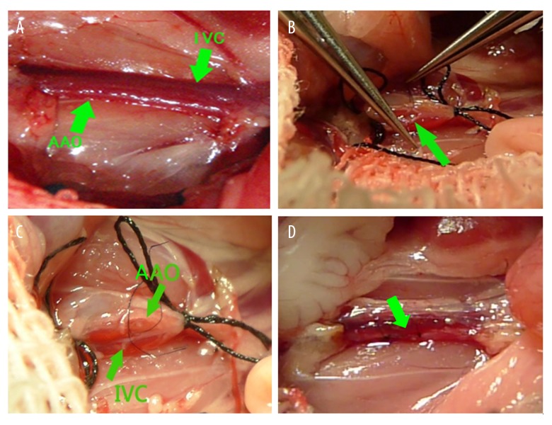
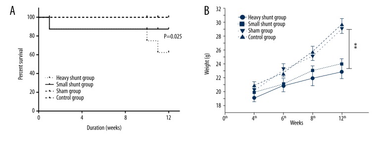
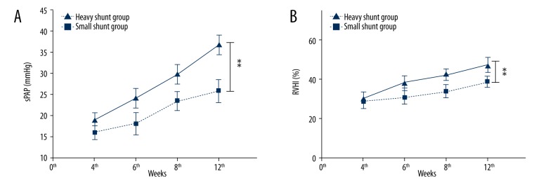
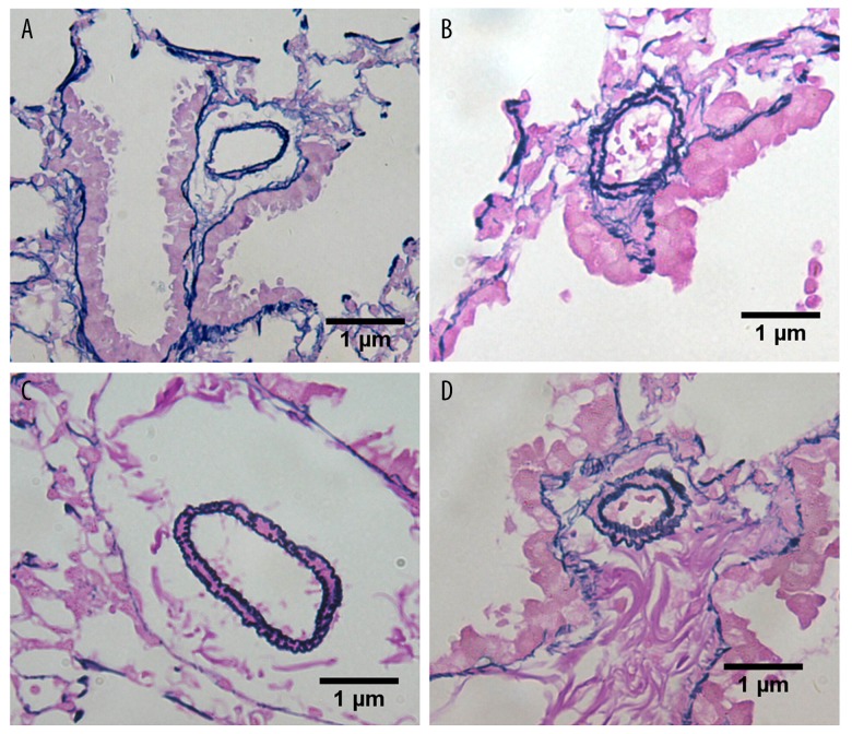
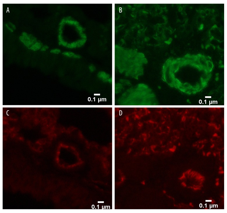
BACKGROUND Establishing a shunt-induced pulmonary arterial hypertension (PAH) model in mice would be of great scientific value, but no such models have been reported to date. Here, we established a shunt-associated PAH in mice to investigate the characteristics of pulmonary vascular remodeling, which provides a new platform for the in-depth study of PAH associated with congenital heart disease (CHD). MATERIAL AND METHODS Eighty mice were randomly divided into the heavy shunt group (n=32), the small shunt group (n=32), the sham operation group (n=8), and the control group (n=8). The septum of the abdominal aorta and inferior vena cava was cut directly to create a heavy abdominal aortocaval shunt. Pulmonary artery pressure, right ventricular hypertrophy index, and lung tissue morphology were evaluated in the 4th, 6th, 8th, and 12th weeks in the shunt groups. RESULTS Shunt-associated PAH by abdominal aortocaval shunt in mice was successfully established. The shunt patency rate was significantly higher in the heavy shunt group. Significant differences were observed between the heavy shunt group and other groups in terms of pulmonary artery pressure and the right ventricular hypertrophy index. Tissue sections revealed a thickened pulmonary intimal layer and muscular layer and stenosis of the lumen in the shunt groups. Immunofluorescent assay results showed significant proliferations of PAH smooth muscle cells and endothelial cells, consistent with the clinical pulmonary vascular remodeling seen in human patients with severe PAH. CONCLUSIONS Shunt-associated PAH established by directly cutting the septum between the abdominal aorta and inferior vena cava is a stable and reliable model for research on PAH associated with CHD.


Conflict of interest statement

Conflicts of interest:

None.

Figures

Figure 1
Figure 1
Surgical procedure of creation of the shunt. (A) The abdominal aorta (AAo) and inferior vena cava (IVC) were tightly linked together; (B) Blood in the abdominal aorta and the inferior vena cava were blocked and a transverse incision were made in the abdominal aorta’s anterior wall to expose the contralateral wall, and then the wall were cut by micro-scissors; (C) Normal saline was injected from the abdominal aorta and the inferior vena cava was filled with normal saline; (D) After the shunt was made, the inferior vena cava pulsed with arterial blood.
Figure 2
Figure 2
Survival and growth in each group. Three mice in the heavy shunt group and 1 in the small shunt group died after the shunt was established. All mice in the sham operation and control groups survived. Mice in the heavy shunt group had higher mortality than those in other groups (P=0.025) (A). Mice in the heavy shunt group had significantly reduced weight at the end of the 8th and 12th weeks compared to mice in the small shunt group (P=0.002 and 0.017, respectively). Mice in both shunt groups had significantly reduced weights at the end of the 6th, 8th, and 12th weeks compared to the control and sham groups (P=0.015) (B). (** indicate P≤0.05).
Figure 3
Figure 3
A comparison of the sPAP and RVHI in the shunt groups. Mice in the heavy shunt group had a significantly higher systolic pulmonary artery pressure (sPAP) at the end of the 12th week compared to that in the small shunt group (36.9±2.2 mmHg vs. 25.9±2.6 mmHg, P=0.015) (A). Mice in both shunt groups had a significantly higher sPAP at the end of the 12th week compared to the control and sham groups (P=0.007). Mice in the heavy shunt group had a significantly higher right ventricular hypertrophy index (RVHI) at the end of the 12th week compared to that in the small shunt group (47.2±3.5% vs. 39.1±2.5%, P=0.037). Mice in both shunt groups had a significantly higher RVHI at the end of the 12th week compared to the control and sham groups (P=0.02) (B). (** Indicates P≤0.05)
Figure 4
Figure 4
Pulmonary vascular remodeling in each group. (A) Pulmonary vascular remodeling in the 8th week (×400) showing mild proliferation of smooth muscle cells in the small shunt group; (B) Pulmonary vascular remodeling in the 8th week (×400) showing moderate proliferation of smooth muscle cells in the heavy shunt group; (C) Pulmonary vascular remodeling in the 12th week (×400) showing moderate proliferation of smooth muscle cells in the small shunt group; and (D) Pulmonary vascular remodeling in the 12th week (×400) showing proliferation of endothelial cells in the heavy shunt group.
Figure 5
Figure 5
Pulmonary vascular remodeling in the 12th week. (A) Pulmonary vascular remodeling in the 12th week (×400) showing proliferation of smooth muscle cells (stained in green) in the small shunt group; (B) Pulmonary vascular remodeling in the 12th week (×400) showing obvious proliferation of smooth muscle cells (stained in green) in the heavy shunt group; (C) Pulmonary vascular remodeling in the 12th week (×400) showing a monolayer of endothelial cells (stained in red) in the small shunt group; and (D) Pulmonary vascular remodeling in the 12th week (×400) showing proliferation of endothelial cells (stained in red) in the heavy shunt group.

Similar articles

Cited by

References

    1. Lowe BS, Therrien J, Ionescu-Ittu R, et al. Diagnosis of pulmonary hypertension in the congenital heart disease adult population impact on outcomes. J Am Coll Cardiol. 2011;58:538–46. - PubMed
    1. Diller GP, Gatzoulis MA. Pulmonary vascular disease in adults with congenital heart disease. Circulation. 2007;115:1039–50. - PubMed
    1. Schulze-Neick I, Deanfield J. Pulmonary arterial hypertension in adults with congenital heart disease: General overview of disease mechanisms. Advances in Pulmonary Hypertension Autumn. 2007;6:121–25.
    1. Canniere DD, Stefanidis C, Brimioulle S, Naeije R. Effects of a chronic aortopulmonary shunt on pulmonary hemodynamics in piglets. J Appl Physiol. 1994;77:1591–96. - PubMed
    1. van Albada ME, Schoemaker RG, Kemna MS, et al. The role of increased pulmonary blood flow in pulmonary arterial hypertension. Eur Respir J. 2005;26:487–93. - PubMed

MeSH terms