Comment
. 2018 May;24(4):408-416.
doi: 10.1038/nm.4512. Epub 2018 Mar 19.

A human monoclonal antibody prevents malaria infection by targeting a new site of vulnerability on the parasite

Affiliations
Comment

A human monoclonal antibody prevents malaria infection by targeting a new site of vulnerability on the parasite

Neville K Kisalu et al. Nat Med. 2018 May.

Erratum in

Abstract

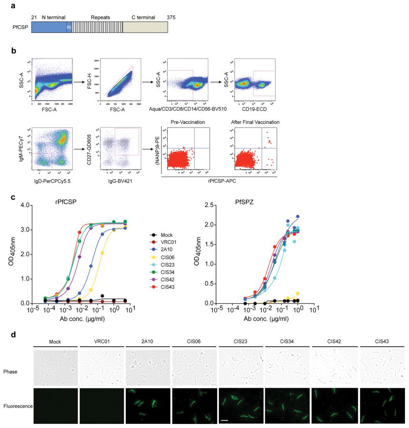
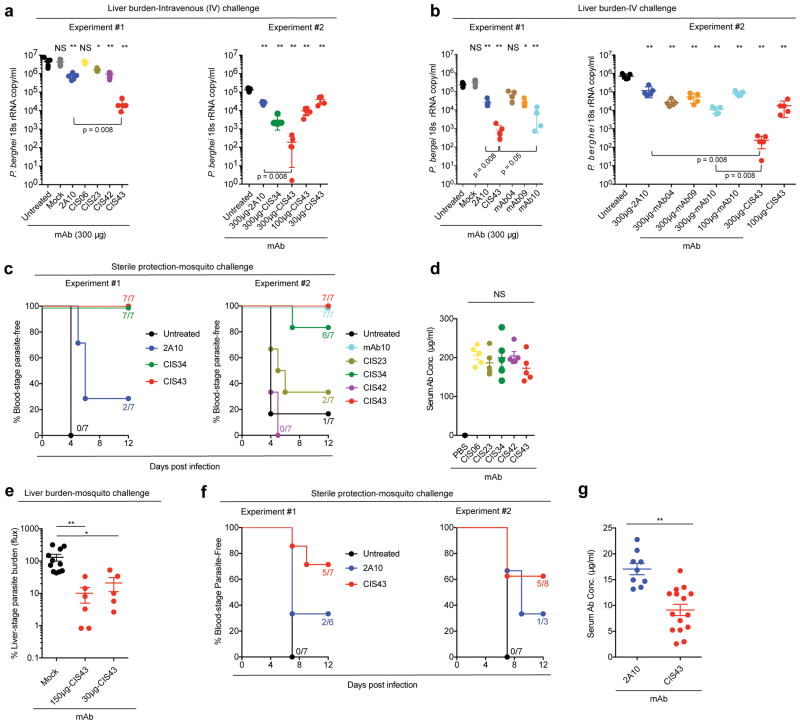
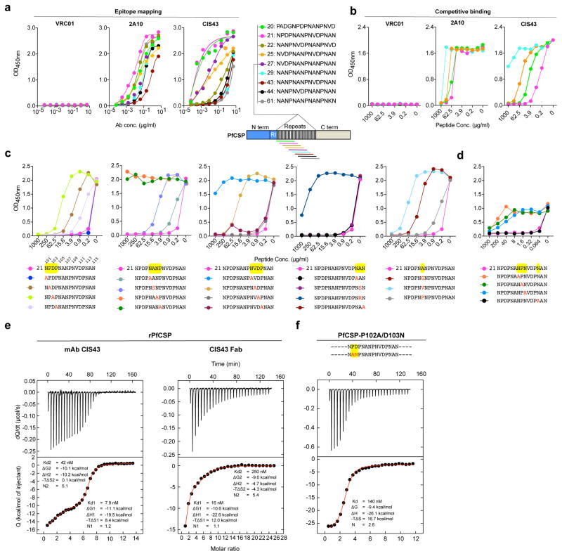
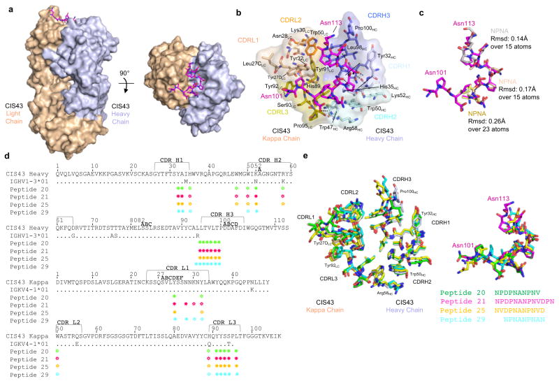
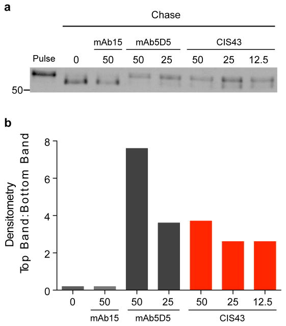
Development of a highly effective vaccine or antibodies for the prevention and ultimately elimination of malaria is urgently needed. Here we report the isolation of a number of human monoclonal antibodies directed against the Plasmodium falciparum (Pf) circumsporozoite protein (PfCSP) from several subjects immunized with an attenuated Pf whole-sporozoite (SPZ) vaccine (Sanaria PfSPZ Vaccine). Passive transfer of one of these antibodies, monoclonal antibody CIS43, conferred high-level, sterile protection in two different mouse models of malaria infection. The affinity and stoichiometry of CIS43 binding to PfCSP indicate that there are two sequential multivalent binding events encompassing the repeat domain. The first binding event is to a unique 'junctional' epitope positioned between the N terminus and the central repeat domain of PfCSP. Moreover, CIS43 prevented proteolytic cleavage of PfCSP on PfSPZ. Analysis of crystal structures of the CIS43 antigen-binding fragment in complex with the junctional epitope determined the molecular interactions of binding, revealed the epitope's conformational flexibility and defined Asn-Pro-Asn (NPN) as the structural repeat motif. The demonstration that CIS43 is highly effective for passive prevention of malaria has potential application for use in travelers, military personnel and elimination campaigns and identifies a new and conserved site of vulnerability on PfCSP for next-generation rational vaccine design.


Conflict of interest statement

Competing financial interests

N.K.C., S.C., B.K.L.S., and S.L.H. are salaried employees of Sanaria Inc., the developer and owner of PfSPZ Vaccine and the sponsor of the clinical trials. In addition, S.L.H. and B.K.L.S. have a financial interest in Sanaria Inc. All other authors declare no competing financial interests.

Figures

Figure 1
Figure 1. Isolation and binding specificity of mAbs from rPfCSP specific-memory B cells
a, Schematic representation of rPfCSP (residues 21–375). Signal (1–21) and anchor (375–397) residues are excluded. The N-, C-terminal, and repeat domains are shown. The conserved region I (RI) is indicated. b, Gating strategy for sorting rPfCSP and (NANP)9 memory B cells. rPfCSP-specific, CD19+ IgG+ CD27+ memory B cells from pre-vaccination or after the 5th (last) vaccination. c, Binding of varying concentrations of mAbs to rPfCSP by ELISA. OD405nm, optical density at 405 nm. d, Binding of mAbs to PfSPZ by ELISA. e, Binding of mAbs to PfSPZ by immunofluorescence assay (IFA). Phase contrast and fluorescence channels are shown. Scale bar, 10 μm. In c–e, Negative controls: Mock, transfection filtrate; VRC01, a human anti-HIV-1 IgG1 isotype control mAb. Positive control: 2A10, mouse anti-PfCSP repeat mAb. Data are representative of two independent experiments (c–e).
Figure 2
Figure 2. Protection against malaria infection by PfCSP mAbs
Protective effect of PfCSP mAbs isolated from PfCSP-specific memory B cells (a) and plasmablasts (b) on liver burden. Following passive transfer of the indicated mAbs, C57BL/6 mice (n=5/group) were challenged intravenously (IV) with chimeric PbSPZ expressing PfCSP (Pb-PfCSP). Liver burden is expressed as Pb 18s ribosomal RNA (rRNA) copies/mL and differences between each antibody and the untreated group was determined using the two-tailed Mann–Whitney test. * p = 0.016, and ** p = 0.008, and NS = not significant. Brackets reflect comparisons between mAb CIS43 and mAb 2A10 or mAb10. c, Sterile protection by PfCSP mAbs following Pb-PfCSP SPZ infection by mosquito bite. C57BL/6 mice challenged with 5 infected mosquitoes following passive transfer (300 ug) of the indicated mAbs. Kaplan Meier curves, analyzed by the log rank test, show frequencies of mice free of parasites as determined by Giemsa staining of blood. Differences between mAbs CIS43, CIS34 and mAb10 as compared to untreated mice were significant (p = 0.0001). d, Serum PfCSP mAb levels one hour after passive transfer of 300 μg the indicated mAbs as determined by ELISA in a separate group of C57BL/6 mice (n = 5/group). Differences in mAb levels were compared for significance using one-way ANOVA, with Dunnett’s correction for multiple comparisons. e, Protective effect of mAb CIS43 on parasite liver burden following Pf infection in FRG-huHep mice with 50 mosquitoes infected with Pf expressing GFP-luciferase. Parasite burden determined 6 days later by bioluminescent imaging (flux). Results were normalized to mice receiving a non-specific IgG (Mock). Mock, n = 12 mice; mAb CIS43 150 and 30 μg, n = 7 and 6 mice, respectively. The Kruskal-Wallis test, with Dunn’s correction was used for multiple comparisons (* p = 0.041 and ** p = 0.001). f, Sterile protection by PfCSP mAbs following PfSPZ infection. Following passive transfer (50 μg) of the indicated mAbs, FRG-huHep mice challenged with 5 infected mosquitoes. Kaplan Meier curves, analyzed by the log rank test, show frequencies of mice free of parasites as determined by Pf 18s rRNA on day 7 and 9. In both experiments mAb CIS43 was significantly more protective than untreated mice (p = 0.0002). g, Serum PfCSP mAb levels in FRG-huHep mice used in f were assessed at the time of challenge. Data were compared for significance using the two-tailed Mann–Whitney test (** p = 0.001). Data are representative of two independent experiments (d and e). Bar denotes geometric mean (panel d, e, f) and median (panel a, b, g). Each dot represents one mouse.
Figure 3
Figure 3. Epitope mapping and ITC analysis of mAb CIS43
a, Binding of mAb CIS43 to overlapping peptides of PfCSP, with specified sequences numbered and color coded 20 – 61 (representing amino acid residues 97–276) by ELISA. Peptides 28–41 and 46–60, consist only of NANP repeats, and are represented by peptide 29. b, Binding of mAb CIS43 to rPfCSP in the presence of varying concentrations of peptides. Peptide color code as in a. c, Binding of mAb CIS43 to rPfCSP in the presence of peptide 21 sequence variants. Wild type peptide 21 sequence with numeric position listed above and mutated residues highlighted in yellow. d, Binding of mAb CIS43 to PfSPZ in the presence of peptide 21 and its sequence variants. e, ITC of mAb CIS43 and CIS43 Fab binding to rPfCSP. Data are representative of two independent experiments (a–e). f, ITC of mAb CIS43 binding to PfCSP mutant, PfCSP(P102A,D103N), with changes in the junctional epitope sequence depicted in red and highlighted in yellow. Upper panels show the output signal, dQ/dt, as a function of time. Lower panels show the integrated heats as a function of the antibody-site/PfCSP molar ratio in the cell. The solid line represents the result from best non-linear leastsquares fit of the data to a binding model that takes into account two sets of sites with different affinities for rPfCSP. Data shown are representative of three independent experiments. Dissociation constant (Kd), changes in Gibbs energy (ΔG) of binding, enthalpy (ΔH) and entropy (−TΔS) and stoichiometry (N) are shown.
Figure 4
Figure 4. Crystal structures of CIS43 Fab in complex with PfCSP peptides
a, Surface representation of CIS43 Fab (light chain in wheat and heavy chain in light blue) with peptide 21 N101PDPNANPNVDPN113 from the repeat region of PfCSP shown in sticks (magenta) and 90° rotation with view down towards the combining sites. b, Details of the interactions between peptide 21 and CIS43 Fab. The peptide 21 is shown in magenta as sticks representation, the CIS43 epitope is shown as sticks and semi-transparent surface and residues are colored based on the CDR regions, with light chain in shades of wheat and heavy chain in shades of light blue. c, Stick representation of peptide 21 shown in magenta in the conformation bound to CIS43 with superposition of three type-I β-turn NPNA repeat structures of CSP as described by Ghasparian et al. Each NPNA repeat is labeled for clarity and shown in different colors. Root mean square deviation (Angstroms, Å) is indicated over the total number of atoms used in the alignment. d, Sequence of CIS43 Fab following Kabbat numbering and alignment with germline gene. Residues that contact each peptide (green, magenta, yellow and cyan for peptide 20, 21, 25 and 29, respectively) are shown as closed circle for main chain only, open star for side chains only and closed star for both main and side chains. e, Detailed of the interactions of the peptides with CIS43 Fabs. Residues within 5 Å of the peptides are shown as sticks in wheat for the light chain residues and light blue for the heavy chain residues when bound to peptide 21 and as green, yellow and cyan for peptide 20, peptide 25 and peptide 29, respectively. (Right) Superposition of the peptides shown as sticks and colored as in (d) with sequences observed in electron density.
Figure 5
Figure 5. Effect of mAb CIS43 on cleavage of PfCSP
a, Metabolically-labeled Pf sporozoites were kept on ice (Pulse) or chased for 90 min in the absence, or presence of the indicated mAb concentrations (μg/ml). PfCSP was immunoprecipitated from sporozoite lysates and analyzed by SDS-PAGE and autoradiography. Negative control: mAb15 (anti C-terminus PfCSP mAb, Supplementary Fig. 1c). Positive control: mAb5D5 (mouse anti-N terminus PfCSP mAb). Molecular mass is indicated in kilodaltons on the left side of the autoradiograph. b, Densitometry analysis of scanned autoradiograph shown in a. The density ratio of top to bottom PfCSP bands is shown for each chased sample with mAb concentrations shown below in (μg/ml). A ratio of 1 indicates the density of the top and bottom bands is equal. Data are representative of 3 independent experiments (a–b).

Comment in

Comment on

Similar articles

Cited by

References

    1. Menard R, et al. Circumsporozoite protein is required for development of malaria sporozoites in mosquitoes. Nature. 1997;385:336–340. doi: 10.1038/385336a0. - DOI - PubMed
    1. Coppi A, et al. Heparan sulfate proteoglycans provide a signal to Plasmodium sporozoites to stop migrating and productively invade host cells. Cell Host Microbe. 2007;2:316–327. doi: 10.1016/j.chom.2007.10.002. - DOI - PMC - PubMed
    1. Ancsin JB, Kisilevsky R. A binding site for highly sulfated heparan sulfate is identified in the N terminus of the circumsporozoite protein: significance for malarial sporozoite attachment to hepatocytes. J Biol Chem. 2004;279:21824–21832. doi: 10.1074/jbc.M401979200. - DOI - PubMed
    1. Rathore D, Sacci JB, de la Vega P, McCutchan TF. Binding and invasion of liver cells by Plasmodium falciparum sporozoites. Essential involvement of the amino terminus of circumsporozoite protein. J Biol Chem. 2002;277:7092–7098. doi: 10.1074/jbc.M106862200. - DOI - PubMed
    1. Dame JB, et al. Structure of the gene encoding the immunodominant surface antigen on the sporozoite of the human malaria parasite Plasmodium falciparum. Science. 1984;225:593–599. - PubMed

Publication types