

4EHP-independent repression of endogenous mRNAs by the RNA-binding protein GIGYF2
- PMID: 29554310
- PMCID: PMC6009589
- DOI: 10.1093/nar/gky198
4EHP-independent repression of endogenous mRNAs by the RNA-binding protein GIGYF2
Abstract
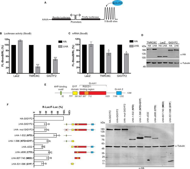
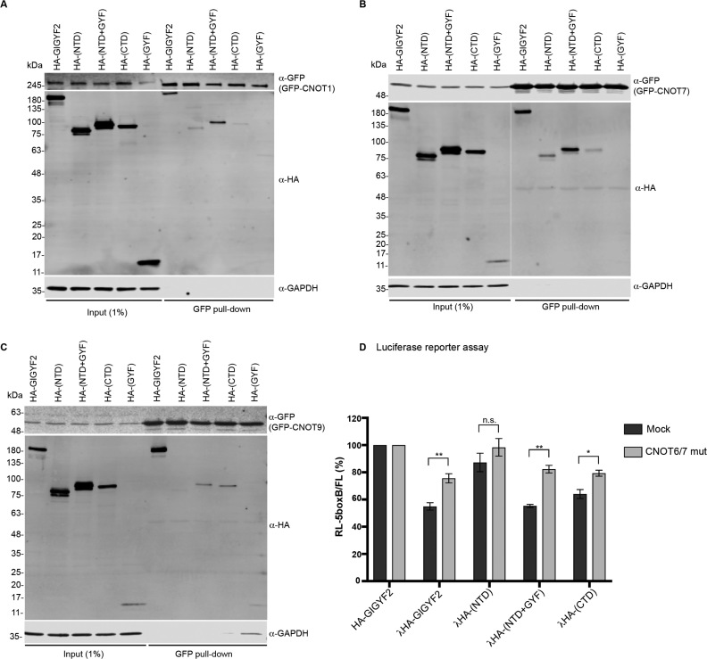
Initially identified as a factor involved in tyrosine kinase receptor signaling, Grb10-interacting GYF protein 2 (GIGYF2) has later been shown to interact with the 5' cap-binding protein 4EHP as part of a translation repression complex, and to mediate post-transcriptional repression of tethered reporter mRNAs. A current model proposes that GIGYF2 is indirectly recruited to mRNAs by specific RNA-binding proteins (RBPs) leading to translation repression through its association with 4EHP. Accordingly, we recently observed that GIGYF2 also interacts with the miRNA-induced silencing complex and probably modulates its translation repression activity. Here we have further investigated how GIGYF2 represses mRNA function. In a tethering reporter assay, we identify three independent domains of GIGYF2 with repressive activity. In this assay, GIGYF2-mediated repression is independent of 4EHP but largely dependent on the CCR4/NOT complex that GIGYF2 recruits through multiple interfaces. Importantly, we show that GIGYF2 is an RBP and identify for the first time endogenous mRNA targets that recapitulate 4EHP-independent repression. Altogether, we propose that GIGYF2 has two distinct mechanisms of repression: one depends on 4EHP binding and mainly affects translation; the other is 4EHP-independent and involves the CCR4/NOT complex and its deadenylation activity.
Figures


Similar articles
-
Direct role for the Drosophila GIGYF protein in 4EHP-mediated mRNA repression.Nucleic Acids Res. 2019 Jul 26;47(13):7035-7048. doi: 10.1093/nar/gkz429. Nucleic Acids Res. 2019. PMID: 31114929 Free PMC article.
-
GIGYF1/2 proteins use auxiliary sequences to selectively bind to 4EHP and repress target mRNA expression.Genes Dev. 2017 Jun 1;31(11):1147-1161. doi: 10.1101/gad.299420.117. Epub 2017 Jul 11. Genes Dev. 2017. PMID: 28698298 Free PMC article.
-
Post-transcriptional gene silencing activity of human GIGYF2.Biochem Biophys Res Commun. 2016 Jul 1;475(3):289-94. doi: 10.1016/j.bbrc.2016.05.022. Epub 2016 May 5. Biochem Biophys Res Commun. 2016. PMID: 27157137
-
GIGYF2: A Multifunctional Regulator at the Crossroads of Gene Expression, mRNA Surveillance, and Human Disease.Cells. 2025 Jul 5;14(13):1032. doi: 10.3390/cells14131032. Cells. 2025. PMID: 40643551 Free PMC article. Review.
-
eIF4E-homologous protein (4EHP): a multifarious cap-binding protein.FEBS J. 2023 Jan;290(2):266-285. doi: 10.1111/febs.16275. Epub 2021 Nov 29. FEBS J. 2023. PMID: 34758096 Review.
Cited by
-
Direct role for the Drosophila GIGYF protein in 4EHP-mediated mRNA repression.Nucleic Acids Res. 2019 Jul 26;47(13):7035-7048. doi: 10.1093/nar/gkz429. Nucleic Acids Res. 2019. PMID: 31114929 Free PMC article.
-
Regulation of the Golgi Apparatus by p38 and JNK Kinases during Cellular Stress Responses.Int J Mol Sci. 2021 Sep 4;22(17):9595. doi: 10.3390/ijms22179595. Int J Mol Sci. 2021. PMID: 34502507 Free PMC article.
-
Repression of mRNA translation initiation by GIGYF1 via disrupting the eIF3-eIF4G1 interaction.Sci Adv. 2024 Jul 19;10(29):eadl5638. doi: 10.1126/sciadv.adl5638. Epub 2024 Jul 17. Sci Adv. 2024. PMID: 39018414 Free PMC article.
-
Molecular basis for GIGYF-TNRC6 complex assembly.RNA. 2023 Jun;29(6):724-734. doi: 10.1261/rna.079596.123. Epub 2023 Feb 28. RNA. 2023. PMID: 36854607 Free PMC article.
-
Genome-Wide Identification of GYF-Domain Encoding Genes in Three Brassica Species and Their Expression Responding to Sclerotinia sclerotiorum in Brassica napus.Genes (Basel). 2023 Jan 15;14(1):224. doi: 10.3390/genes14010224. Genes (Basel). 2023. PMID: 36672966 Free PMC article.
References
-
- Giovannone B., Lee E., Laviola L., Giorgino F., Cleveland K.A., Smith R.J.. Two novel proteins that are linked to insulin-like growth factor (IGF-I) receptors by the Grb10 adapter and modulate IGF-I signaling. J. Biol. Chem. 2003; 278:31564–31573. - PubMed
-
- Ajiro M., Nishidate T., Katagiri T., Nakamura Y.. Critical involvement of RQCD1 in the EGFR-Akt pathway in mammary carcinogenesis. Int. J. Oncol. 2010; 37:1085–1093. - PubMed
-
- Higashi S., Iseki E., Minegishi M., Togo T., Kabuta T., Wada K.. GIGYF2 is present in endosomal compartments in the mammalian brains and enhances IGF-1-induced ERK1/2 activation. J. Neurochem. 2010; 115:423–437. - PubMed
-
- Tan E.K., Schapira A.H.. Summary of GIGYF2 studies in Parkinson's disease: the burden of proof. Eur. J. Neurol. 2010; 17:175–176. - PubMed
Publication types
MeSH terms
Substances
LinkOut - more resources
Full Text Sources
Other Literature Sources
Molecular Biology Databases
Research Materials
Miscellaneous
