

Can Dynamic MRI Be Used to Accurately Identify Velopharyngeal Closure Patterns?
- PMID: 29554453
- PMCID: PMC6463292
- DOI: 10.1177/1055665617735998
Can Dynamic MRI Be Used to Accurately Identify Velopharyngeal Closure Patterns?
Abstract
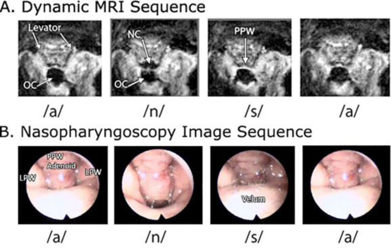
Background: Dynamic magnetic resonance imaging (MRI) has been proposed as a non-invasive, child-friendly, reproducible, and repeatable imaging method providing a 3-dimensional view of the velopharyngeal structures and function during speech. However, the value of dynamic MRI as compared to imaging methods such as nasopharyngoscopy is not well understood. The aim of this study was to compare the ability of nasopharyngoscopy and dynamic MRI to accurately identify velopharyngeal closure patterns among adults without cleft palate.
Methods: Participants included 34 healthy adults with normal anatomy between 19 and 33 years of age (mean = 23 years; SD = 4.1 years). Participants underwent dynamic MRI and nasopharyngoscopy studies and comparisons were performed to determine the intra- and inter-rater reliability for accurately determining closure pattern. The MRI acquisition was a dynamic acquisition of a 2D plane.
Results: Strong inter- (κ = .824; P < .001) and intra-rater (Rater 1: κ = 0.879, P < .001, 94% agreement between ratings; Rater 2 with 100% agreement) agreement was observed for the identification of closure pattern using nasopharyngoscopy. Inter-rater agreement for ratings using MRI demonstrated moderate agreement (κ = .489; P < .004). Examining point agreement revealed only 27 of the 33 ratings of MRI showed agreement (80%).
Conclusion: This demonstrates that inter-rater reliability for determining closure patterns from nasopharyngoscopy is good; however, ratings using MRI was less reliable at determining closure patterns. It is likely that future improvements in dynamic imaging with MRI to enable 3D visualizations are needed for improved diagnostic accuracy for assessing velopharyngeal closure patterns.
Keywords: magnetic resonance imaging; nasopharyngoscopy; velopharyngeal function.
Figures


References
-
- Armour A, Fischbach S, Klaiman P, Fisher DM. Does velopharyngeal closure pattern affect the success of pharyngeal flap pharyngoplasty? Plast Reconstr Surg. 2005;115:45–52. - PubMed
-
- Beer AJ, Hellerhoff P, Zimmermann A, Mady K, Sader R, Rummeny EJ, Hannig C. Dynamic near-real-time magnetic resonance imaging for analyzing the velopharyngeal closure in comparison with videofluoroscopy. J Magn Reson Imaging. 2004;20:791–797. - PubMed
-
- Bicknell S, McFadden LR, Curran JB. Frequency of pharyngoplasty after primary repair of cleft palate. J Can Dent Assoc. 2002;68(11):688–692. . - PubMed
-
- Cable BB, Canady JW. The endoscopically assisted pharyngeal flap. Cleft Palate Craniofac J. 2003;40:114–115. - PubMed
-
- Ettema SL, Kuehn DP, Perlman AL, Alperin N. Magnetic resonance imaging of the levator veli palatini muscle during speech. Cleft Palate Craniofac J. 2002;39:130–144. - PubMed
Publication types
MeSH terms
Grants and funding
LinkOut - more resources
Full Text Sources
Other Literature Sources
Medical
