. 2018 Jul:305:1-12.
doi: 10.1016/j.expneurol.2018.02.015. Epub 2018 Mar 16.

Impaired social behaviors and minimized oxytocin signaling of the adult mice deficient in the N-methyl-d-aspartate receptor GluN3A subunit

Affiliations

Impaired social behaviors and minimized oxytocin signaling of the adult mice deficient in the N-methyl-d-aspartate receptor GluN3A subunit

Jin Hwan Lee et al. Exp Neurol. 2018 Jul.

Abstract

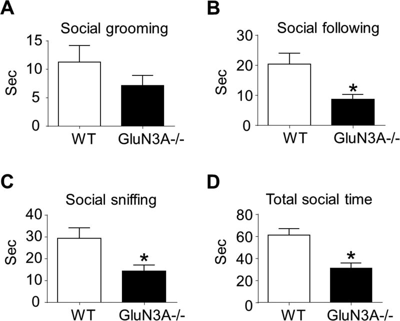
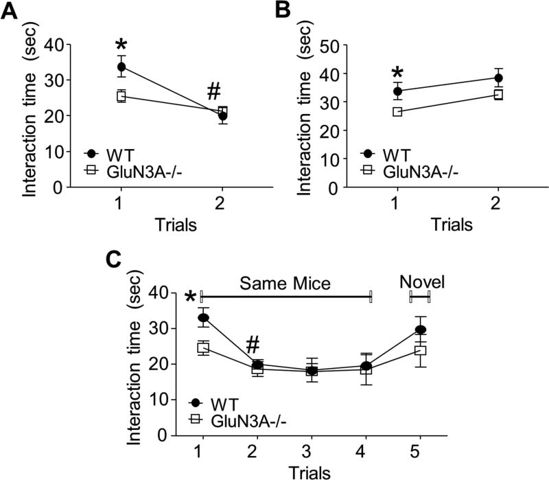
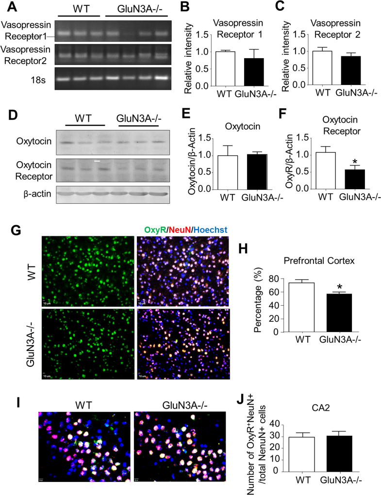
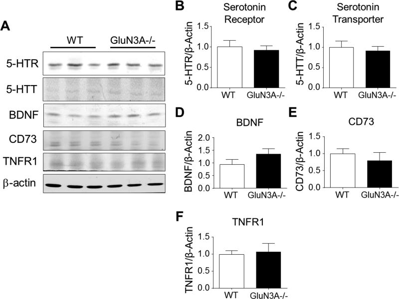
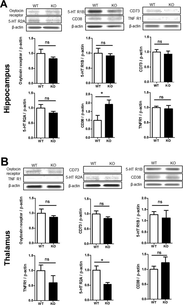
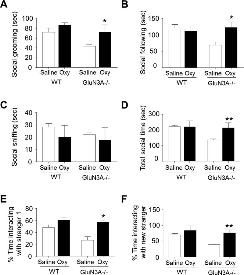
The N-methyl-d-aspartate receptor (NMDAR) has been implicated in the pathophysiology of neurological diseases, such as schizophrenia, autism spectrum disorders (ASD), and Alzheimer's disease (AD), whose unique clinical hallmark is a constellation of impaired social and/or cognitive behaviors. GluN3A (NR3A) is a unique inhibitory subunit in the NMDAR complex. The role of GluN3A in social behavioral activities is obscure. In this study, we sought to evaluate altered social activities in adult GluN3A knockout (KO) mice. GluN3A KO mice spent less time in reciprocal social interaction in the social interaction test compared to wild-type (WT) mice. A social approach test using a three-chamber system confirmed that mice lacking GluN3A had lower sociability and did not exhibit a preference for social novelty. GluN3A KO mice displayed abnormal food preference in the social transmission of food preference task and low social interaction activity in the five-trial social memory test, but without social memory deficits. Using a home cage monitoring system, we observed reduced social grooming behavior in GluN3A KO mice. Signaling genes that might mediate the altered social behaviors were examined in the prefrontal cortex, hippocampus, and thalamus. Among nine genes examined, the expression of the oxytocin receptor was significantly lower in the prefrontal cortex of GluN3A KO mice than that in WT mice. Oxytocin treatment rescued social activity deficits in GluN3A KO mice. These findings support a novel idea that a chronic state of moderate increases in NMDAR activities may lead to downregulation of the oxytocin signaling and impaired behavioral activities that are seen in psychiatric/neurodegenerative disorders.

Keywords: GluN3A; NR3A; Oxytocin signaling; Social behaviors.


Conflict of interest statement

Conflict of Interest

Authors declare no conflict of interest associated with this investigation.

Figures

Figure 1
Figure 1. GluN3A KO mice show deficits in reciprocal social interaction
A social interaction test was performed to reveal the effect of GluN3A on general social activity. A–D. We scored social sniffing, following, grooming, and total social time to quantify reciprocal social interaction. In this test, the trend of reduced social grooming in GluN3A mice was not statistically significant from WT mice. However, social following and social sniffing time, as well as the total social time, were significantly and markedly reduced in GluN3A KO mice compared to WT mice. Paired t test for A – C; Two-way ANOVA followed by Bonferroni correction: F(12,12)=1.516; *P<0.05 vs. WT; n=13 animals per group.
Figure 2
Figure 2. GluN3A KO mice show differences in preference for social novelty in the social approach test and in the social activity test
A social approach test using a three-chamber system was performed to determine whether the deletion of GluN3A could affect social approach, especially sociability and social novelty. A. In the first phase (sociability) of the social approach test, WT mice preferred to stay with the stranger mouse, while GluN3A KO mice spent significantly less time in a chamber containing a stranger mouse. *P<0.05 vs. WT; n=13 per group. B. In the second phase (social novelty) of the social approach test, again, WT mice spent most time with the novel stranger in the third chamber, but the time spent by GluN3A KO mice was significantly less compared to WT mice. *P<0.05 vs. WT; n=13 per group. C. The social transmission of food preference task was a measure of social activity. The carton diagrams on the left show the contact time of a demonstrator mouse (black) with cued food (black) and interaction with an observer mouse; the third diagram shows the observer mouse was placed with both cued and non-cued food in a cage 24 hours after staying with the demonstrator mouse. The bar graph summarized the consumed cued and non-cued food in the third cage. WT mice illustrated a substantial preference on cued food, apparently influenced by the demonstrator. On the contrary, GluN3A KO mice showed no significant difference in eating cued and non-cued food. One-way ANOVA followed by Bonferroni correction; F(1, 40)=15, 11, p<0.05, n=14 per group.
Figure 3
Figure 3. GluN3A KO mice exhibited normal social memory function
Direct interaction and five-trial social memory tests were performed to investigate the effect of GluN3A on social memory. A and B. Setup of the social direct interaction test using the same (A) or different (B) mice in the two trials. A. WT mice showed proper habituation behavior with a decreased interaction time in trial 2, which is suggestive of intact social memory. Although GluN3A KO mice displayed relatively decreased habituation across two trials compared to WT mice, as indicated by their decreased interaction time during the second exposure, but the inter-trial difference was not significant (P=0.1157). One-way ANOVA; F(1,10)=5.725, *P<0.05 vs. WT, #P<0.05 vs. same group trial 1; n=7 animals per group. B. When subject mice were exposed to a novel mouse, both groups explored new stimulus mice similarly. One-way ANOVA; F(1.10)=0.06590, *P<0.05 vs. WT; n=7 per group. C. Five-trial social memory assay. Although GluN3A KO mice showed decreased interaction times, both WT and GluN3A KO mice habituated to the same mice during four trials and dishabituated to novel mice. Two-way ANOVA, F(4.40)=0.6176, *P<0.05 vs. WT, #P<0.05 vs. same group; n=7 per group. Student’s two-tailed t-test was applied.
Figure 4
Figure 4. Deletion of GluN3A leads to abnormal development of oxytocin receptor
We next performed RT-PCR analysis, Western blotting, and immunohistochemistry to investigate the possibility that the deletion of GluN3A could alter the development of the oxytocin system and vasopressin system. A. RT-PCR analysis of vasopressin (AVP) receptors 1 and 2 in the prefrontal cortex of WT and GluN3A KO mice. B and C. Quantification of relative intensity (normalized to 18S ribosomal RNA control) of AVP receptors 1 and 2 in the prefrontal cortex. Student t test; n=6 per group. D. Western blotting of oxytocin and oxytocin receptor in the prefrontal cortex from WT and GluN3A KO mice. E and F. Quantification of the optic density (normalized to β-actin control) of oxytocin and oxytocin receptor in the prefrontal cortex. Student t test; *P<0.05 vs. WT; n=6 per group. G. Immunohistochemistry analysis revealed that oxytocin receptor-positive cells were completely co-labeled with NeuN positive cells in the prefrontal cortex. H. In the prefrontal cortex, the ratio of oxytocin receptor (green)-NeuN (red) double-positive cells to total NeuN-positive cells was lower in GluN3A KO mice as compared to WT mice. Student t test; *P<0.05 vs. WT; n=4–5 per group. I. Immunohistochemistry analysis revealed oxytocin receptor-positive cells in CA2 area of hippocampus. Scale bars = 50 µM. J. The number of oxytocin receptor-NeuN double-positive cells was similar in GluN3A KO mice as compared to WT mice. Student t test; *P<0.05 vs. WT; n=4–5 per group.
Figure 5
Figure 5. GluN3A had no effect on the development of serotonin regulation, BDNF, CD73, and TNFR1
We investigated the possibility that the deletion of GluN3A could altered social behavior-associated genes such as serotonin receptor (5-HTR), serotonin transporter (5-HTT), BDNF, CD73, and TNFR1. A. Western blotting of serotonin receptor, serotonin transporter, BDNF, CD73, and TNFR1 in the prefrontal cortex from WT and GluN3A KO mice. B-F. Quantification of the optic density (normalized to β-actin control) of 5-HTR, 5-HTT, BDNF, CD73, and TNFR1 in the prefrontal cortex. The expression of these genes was also unchanged in GluN3A KO mice. N=6 per group.
Figure 6
Figure 6. Expression of social behavior genes in the hippocampus and thalamus
Western blot analysis was performed on brain tissues from the hippocampus and thalamus areas of WT and GluN3A KO mice. A. Western blot images and quantification of the band intensity compared to the basal level. The protein level of oxytocin receptor, serotonin receptors 5-HTR2A, 5-HTR1B, TNFR1 and CD73 showed no statistically difference between WT and KO mice. The level of CD38, however, was significantly higher in the KO hippocampus. N=6, Student t test; * p<0.05, F=1.299. B. The above assay was repeated in the thalamus. All measured protein expressions were similar between WT and KO mice, expect the 5-HTR2A level was significantly lower in this brain region. N=6; Student t test; * p<0.05, F=5.650.
Figure 7
Figure 7. Oxytocin treatment rescues impaired social behaviors in GluN3A KO mice
Oxytocin (Oxy; 10 mg/kg) was intraperitoneally administered once per day for 7 days in adult WT and GluN3A KO mice. A–D. In the social interaction test, oxytocin treatment rescued the social deficits in social grooming, social following and, consequently, increased the total social time of GluN3A KO mice. One-way ANOVAP; F(1,16)=0.4797 (A), F(1,16)=5.010 (B), F(1,16)=0.2161 (C), F(1,16)=10.21 (D), *P<0.05 vs. saline-treated GluN3A KO mice, **P<0.01 vs. saline-treated GluN3A KO mice; n=5 per group. E. In the sociability test of three chamber system, oxytocin-treated GluN3A KO mice spent significantly more time in the chamber containing a stranger mouse. One-way ANOVA, F(1,8)=3.809, *P<0.05 vs. saline-treated GluN3A KO mice; n=5 per group. F. In the social novelty test, GluN3A KO mice showed social deficits of reduced interaction with a stranger while oxytocin treatment completely corrected this problem to the WT control level. One-way ANOVA, F(1,8)=0.8830; **P<0.01 vs. saline-treated GluN3A KO; n=5 per group.

Similar articles

Cited by

References

    1. Akillioglu K, Binokay S, Kocahan S. The effect of neonatal N-methyl-D-aspartate receptor blockade on exploratory and anxiety-like behaviors in adult BALB/c and C57BL/6 mice. Behav Brain Res. 2012;233:157–161. - PubMed
    1. Al-Hallaq RA, Jarabek BR, Fu Z, Vicini S, Wolfe BB, Yasuda RP. Association of NR3A with the N-methyl-D-aspartate receptor NR1 and NR2 subunits. Mol Pharmacol. 2002;62:1119–1127. - PubMed
    1. Alvarez P, Wendelken L, Eichenbaum H. Hippocampal formation lesions impair performance in an odor-odor association task independently of spatial context. Neurobiol Learn Mem. 2002;78:470–476. - PubMed
    1. Anderson SW, Bechara A, Damasio H, Tranel D, Damasio AR. Impairment of social and moral behavior related to early damage in human prefrontal cortex. Nature Neurosci. 1999;2:1032–1037. - PubMed
    1. Bales KL, Solomon M, Jacob S, Crawley JN, Silverman JL, Larke RH, Sahagun E, Puhger KR, Pride MC, Mendoza SP. Long-term exposure to intranasal oxytocin in a mouse autism model. Transl Psychiatry. 2014;4:e480. - PMC - PubMed

Publication types