Review
. 2018 Aug:51:44-52.
doi: 10.1016/j.sbi.2018.03.009. Epub 2018 Mar 16.

Structural biology of G protein-coupled receptors: new opportunities from XFELs and cryoEM

Affiliations
Review

Structural biology of G protein-coupled receptors: new opportunities from XFELs and cryoEM

Andrii Ishchenko et al. Curr Opin Struct Biol. 2018 Aug.

Abstract

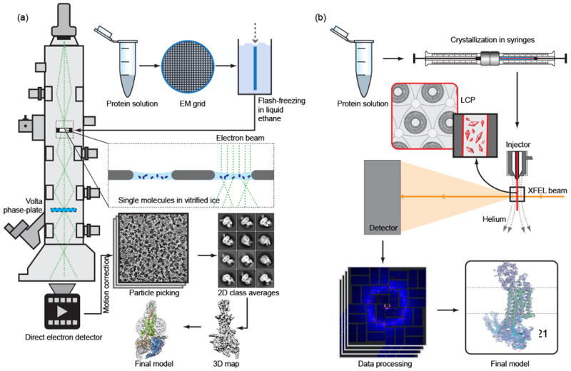
G protein-coupled receptors mediate cell signaling and regulate the majority of sensory and physiological processes in the human body. Recent breakthroughs in cryo-electron microscopy and X-ray free electron lasers have accelerated structural studies of difficult-to-crystallize receptors and their signaling complexes, and have opened up new opportunities in understanding conformational dynamics and visualizing the process of receptor activation with unprecedented spatial and temporal resolution. Here, we summarize major milestones and challenges associated with the application of these techniques and outline future directions in their development with a focus on membrane protein structural biology.


Figures

Figure 1
Figure 1. Schematic diagrams of cryoEM and LCP-SFX experiments
(a) For cryoEM, the purified monodisperse protein solution is deposited on EM grids, blotted and flash-frozen in liquid ethane. The grids are then cryo-transferred into the electron microscope and thousands of images are collected by a direct-electron detector. After performing motion correction, individual particles are picked and 2D classification and 3D classification is applied. Finally, a 3D map is reconstructed, which is used to fit and refine a structure model. Images from Ref. have been re-used in this illustration with permission from Macmillan Publishers Ltd. (b) For LCP-SFX, purified protein is reconstituted in LCP, and crystallization is set up in syringes. After microcrystals have grown, samples from several syringes are consolidated and transferred into a viscous media injector. Tens to hundreds of thousands of diffraction images are collected from microcrystals intersecting the XFEL beam in random orientations. After data processing with specialized software, the structure is solved and refined by standard crystallographic approaches.
Figure 2
Figure 2. Timeline of major milestones (right) and published GPCR structures (left) achieved with XFELs and cryoEM
Figure 3
Figure 3. Examples of GPCR structures determined by LCP-SFX and cryoEM
(a) Structure of the rhodopsin-arrestin complex (PDB ID 5W0P) solved using LCP-SFX at 3.0 Å (anisotropic) resolution. (b) The 1.9 Å resolution structure of the adenosine A2A receptor (PDB ID 5K2C) solved by the sulfur single anomalous dispersion (S-SAD) method using data collected by LCP-SFX. (c) The 4.1 Å resolution structure of the GLP-1 receptor in complex with its native agonist peptide, Gs protein and a stabilizing nanobody (PDB ID 5VAI) obtained by single-molecule cryoEM. All structures are shown in cartoon representation with fusion partners and the nanobody colored in gray. Transparent surface is shown for signaling partners, arrestin, and Gs protein. Ligands, lipids and ions are shown as van der Waals spheres, water molecules as small red spheres. The membrane boundaries are shown as red (extracellular) and blue (intracellular) lines. (d) – (f) Electron density of transmembrane helix III is shown as a blue mesh for corresponding structures in (a) – (c). In (d) and (e) 2Fo-Fc density is contoured at 1s level, in (f) the density is contoured at the authors’ recommended level of 0.055 [44].

Similar articles

Cited by

References

    1. Chun E, Thompson AA, Liu W, Roth CB, Griffith MT, Katritch V, Kunken J, Xu F, Cherezov V, Hanson MA, et al. Fusion partner toolchest for the stabilization and crystallization of G protein-coupled receptors. Structure. 2012;20:967–76. - PMC - PubMed
    1. Rosenbaum DM, Cherezov V, Hanson MA, Rasmussen SGF, Thian FS, Kobilka TS, Choi H-J, Yao X-J, Weis WI, Stevens RC, et al. GPCR Engineering Yields High-Resolution Structural Insights into β2-Adrenergic Receptor Function. Science. 2007;318:1266–1273. - PubMed
    1. Serrano-Vega MJ, Magnani F, Shibata Y, Tate CG. Conformational thermostabilization of the β1-adrenergic receptor in a detergent-resistant form. Proc Natl Acad Sci USA. 2008;105:877–882. - PMC - PubMed
    1. Caffrey M, Cherezov V. Crystallizing membrane proteins using lipidic mesophases. Nat Protoc. 2009;4:706–31. - PMC - PubMed
    1. Cherezov V. Lipidic cubic phase technologies for membrane protein structural studies. Curr Opin Struct Biol. 2011;21:559–66. - PMC - PubMed

Publication types

MeSH terms

Substances

LinkOut - more resources