. 2018 Mar 20;37(1):62.
doi: 10.1186/s13046-018-0732-4.

Induction of apoptosis in imatinib sensitive and resistant chronic myeloid leukemia cells by efficient disruption of bcr-abl oncogene with zinc finger nucleases

Affiliations

Induction of apoptosis in imatinib sensitive and resistant chronic myeloid leukemia cells by efficient disruption of bcr-abl oncogene with zinc finger nucleases

Ningshu Huang et al. J Exp Clin Cancer Res. .

Abstract

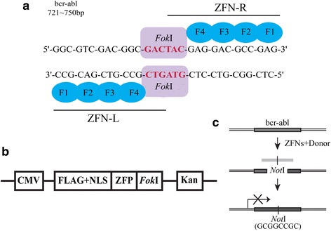
Background: The bcr-abl fusion gene is the pathological origin of chronic myeloid leukemia (CML) and plays a critical role in the resistance of imatinib. Thus, bcr-abl disruption-based novel therapeutic strategy may warrant exploration. In our study, we were surprised to find that the characteristics of bcr-abl sequences met the design requirements of zinc finger nucleases (ZFNs).

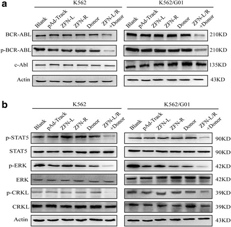
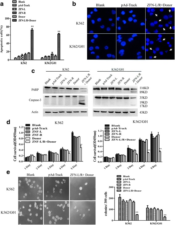
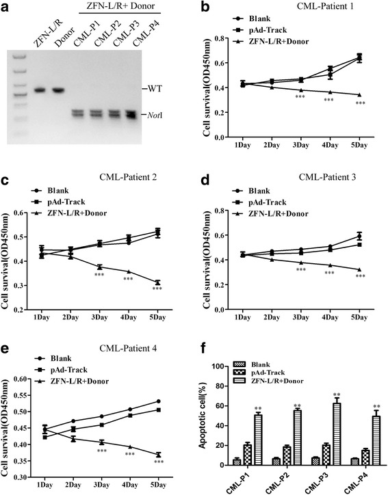
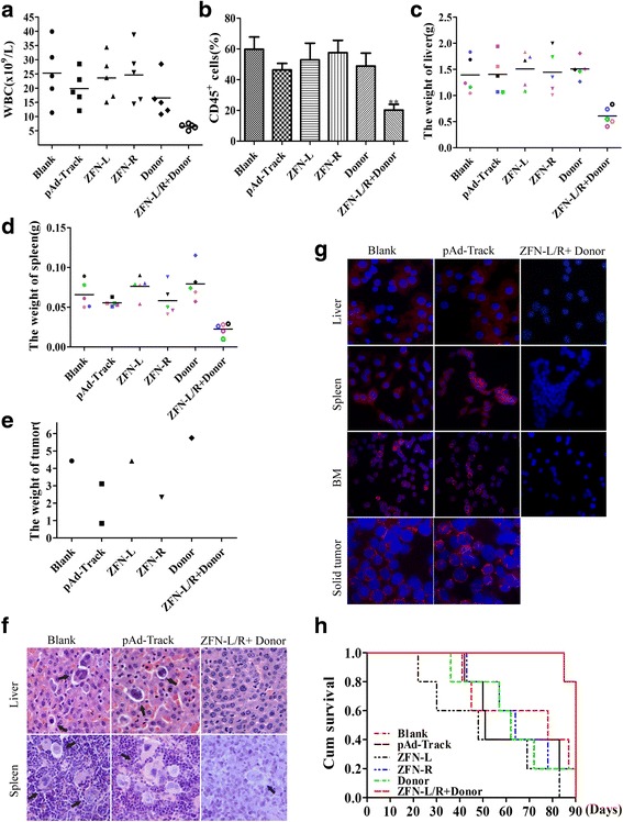
Methods: We constructed the ZFNs targeting bcr-abl with high specificity through simple modular assembly approach. Western blotting was conducted to detect the expression of BCR-ABL and phosphorylation of its downstream STAT5, ERK and CRKL in CML cells. CCK8 assay, colony-forming assay and flow cytometry (FCM) were used to evaluate the effect of the ZFNs on the viablity and apoptosis of CML cells and CML CD34+ cells. Moreover, mice model was used to determine the ability of ZFNs in disrupting the leukemogenesis of bcr-abl in vivo.

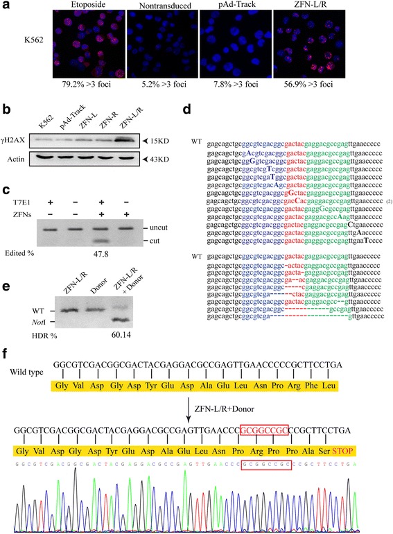
Results: The ZFNs skillfully mediated 8-base NotI enzyme cutting site addition in bcr-abl gene of imatinib sensitive and resistant CML cells by homology-directed repair (HDR), which led to a stop codon and terminated the translation of BCR-ABL protein. As expected, the disruption of bcr-abl gene induced cell apoptosis and inhibited cell proliferation. Notably, we obtained similar result in CD34+ cells from CML patients. Moreover, the ZFNs significantly reduced the oncogenicity of CML cells in mice.

Conclusion: These results reveal that the bcr-abl gene disruption based on ZFNs may provide a treatment choice for imatinib resistant or intolerant CML patients.

Keywords: Bcr-abl; Chronic myeloid leukemia; Homology-directed repair; Oncogenicity; Zinc finger nucleases.


Conflict of interest statement

Ethics approval and consent to participate

All samples were collected with informed consent and all the experiment were approved by the ethical committee. All animal experiments were in accordance with the National Institutes of Health guide for the care and use of Laboratory animals (NIH Publications No. 8023, revised 1978) and were conducted with the approval of the Biomedical Ethics Committee of Chongqing Medical University.

Consent for publication

Not applicable.

Competing interests

The authors declare that they have no competing interests.

Publisher’s Note

Springer Nature remains neutral with regard to jurisdictional claims in published maps and institutional affiliations.

Figures

Fig. 1
Fig. 1
ZFNs were designed to target bcr-abl gene and induce gene modification. a Targeted sequence of ZFNs on bcr-abl gene. ZFN designed to cut exon 1 of bcr-abl gene and consisted of four fingers ZFP and a FokI endonuclease. Together the “left hand” (ZFN-L) and “right hand” (ZFN-R) work as dimers to induce a specific DSB. b The structure of pAd-Track-ZFN vector. ZFP fused to FokI endonuclease, a nuclear localization signal (NLS) and FLAG tag. The expression of Kanomycin resistance gene (Kan) was regulated by CMV promoter. c Sketch of the donor construct and HDR detection scheme. Cleavage of bcr-abl gene created a substrate for HDR, which may use the donor DNA fragment containing a NotI site as a repair template. The introduction of NotI site, which involved 8-bp, may result in termination of translation
Fig. 2
Fig. 2
ZFNs induced gene editing of bcr-abl gene. a K562 cells were treated with 1 μM etoposide (positive control), pAd-Track or ZFN-L/R for 48 h and ZFNs-induced DSBs were detected by 53BP1 immunostaining. Nontransduced cells as negative control. The rate of cells containing more than 3 foci was shown beneath each panel. b K562 cells were transfected with pAd-Track, ZFN-L, ZFN-R plasmids separately or together of ZFN-L and ZFN-R (ZFN-L/R). The amount of γH2AX in each group was quantified by western blot. The arrows indicate the marker proteins. c ZFN-mediated gene editing revealed by T7E1 assay and the results indicated by agarose gel eletrophoresis. The bcr-abl was subjected to digestion with T7E1 to confirm the exist of insertions/deletions. Gene modification was only detected in cells transfected with ZFNs shown as ‘cut’ bands. d The genomic ZFNs target site in K562 cells was sequenced. The result showed the ZFN-induced insertions and deletions around the target region of bcr-abl. e The bcr-abl gene editing efficiency was quantified by NotI restriction enzyme. The genomic DNA of cells transfected with ZFN-L/R, Donor individually or together was extracted and amplified by PCR, then treated with NotI restriction enzyme. “WT” indicates the position of wild type PCR product and “NotI” indicates the position of the fragments generated by NotI digestion. Numbers below the lanes with NotI fragment indicate the rate of PCR product modification. f In silico analysis of sequence of ZFN-L/R and donor treated cells. The result showed the 8-bp (GCGGCCGC) insertion lead to a stop codon and a termination of translation
Fig. 3
Fig. 3
Effect of ZFNs on the expression of BCR-ABL and its downstream signaling pathways. K562 and K562/G01 cells were transfected with pAd-Track, ZFN-L, ZFN-R, Donor or ZFN-L/R and donor. Protein were collected after 48 h for western blotting analysis. a Western blot results showed that ZFN-L/R and donor reduced the expression of BCR-ABL and p-BCR-ABL. b The activity of p-STAT5, p-ERK and p-CRKL were decreased in the group treated with ZFN-L/R and donor
Fig. 4
Fig. 4
ZFNs induces apoptosis and inhibits proliferation of CML cells. K562 and K562/G01 cells were respectively nucleofected with pAd-Track, ZFN-L, ZFN-R, Donor or ZFN-L/R and donor. a The percentage of cell apoptosis was detected by flow cytometry. b Morphologic changes of apoptotic cells were detected by DAPI stain. The arrows indicate the prominent apoptotic morphology. c Cleavage of PARP and Caspase-3 was detected by western blot. d The effect of ZFNs on CML cell proliferation was assessed by CCK-8 assay. e Treated cells were plated in 24-well plates with methylcellulose and colonies were counted after 2 weeks. Data are expressed as the means ± SD. **P < 0.01 vs. Controls
Fig. 5
Fig. 5
ZFNs induces apoptosis and inhibits proliferation of CD34+ cells from CML patients. CML CD34+ cells were collected from patients and treated with ZFN-L/R and Donor plasmid, respectively or together. a The editing efficiency of ZFNs on bcr-abl gene editing was quantified by NotI restriction enzyme. CML-P1, CML-P2, CML-P3 and CML-P4 are abbreviations for CML-Patient 1, CML-Patient 2, CML-Patient 3 and CML-Patient 4 respectively. Cell viability was assessed via CCK8 assay (b), (c), (d), (e) and the percentage of apoptotic cells was determined by FCM (f). The data are shown as the mean ± SD. **P < 0.01 vs. Controls and ***P < 0.001 vs. Controls
Fig. 6
Fig. 6
ZFNs impairs the pathogenecity of bcr-abl in mice. a The maximum of WBC counts of each mouse in Blank, pAd-Track, ZFN-L, ZFN-R, Donor or ZFN-L/R and donor groups were recorded. b The rate of human CD45+ cells in murine bone marrow were detected by flow cytometry. c, d Mean liver or spleen weight of mice in different group was quantified. e The weight of solid tumor for each groups were recorded. f Infiltration of liver or spleen in groups of Blank, pAd-Trackand or ZFN-L/R + donor was analysed by H&E. The arrows indicate the infiltrating leukemic cells. g Detection of BCR-ABL protein by immunofluorescent assay infiltrating. h Survival curves were measured by Kaplan-Meier methods

Similar articles

Cited by

References

    1. Druker BJ. Translation of the Philadelphia chromosome into therapy for CML. Blood. 2008;112:4808–4817. doi: 10.1182/blood-2008-07-077958. - DOI - PubMed
    1. Heisterkamp N, Stephenson JR, Groffen J, Hansen PF, de Klein A, Bartram CR, Grosveld G. Localization of the c-ab1 oncogene adjacent to a translocation break point in chronic myelocytic leukaemia. Nature. 1983;306:239–242. doi: 10.1038/306239a0. - DOI - PubMed
    1. Ben-Neriah Y, Daley GQ, Mes-Masson AM, Witte ON, Baltimore D. The chronic myelogenous leukemia-specific P210 protein is the product of the bcr/abl hybrid gene. Science. 1986;233:212–214. doi: 10.1126/science.3460176. - DOI - PubMed
    1. Lugo TG, Pendergast AM, Muller AJ, Witte ON. Tyrosine kinase activity and transformation potency of bcr-abl oncogene products. Science. 1990;247:1079–1082. doi: 10.1126/science.2408149. - DOI - PubMed
    1. Steelman LS, Pohnert SC, Shelton JG, Franklin RA, Bertrand FE, McCubrey JA. JAK/STAT, Raf/MEK/ERK, PI3K/Akt and BCR-ABL in cell cycle progression and leukemogenesis. Leukemia. 2004;18:189–218. doi: 10.1038/sj.leu.2403241. - DOI - PubMed

MeSH terms