. 2018 Apr 3;115(14):E3305-E3312.
doi: 10.1073/pnas.1715625115. Epub 2018 Mar 19.

Flexible egocentric and allocentric representations of heading signals in parietal cortex

Affiliations

Flexible egocentric and allocentric representations of heading signals in parietal cortex

Xiaodong Chen et al. Proc Natl Acad Sci U S A. .

Abstract

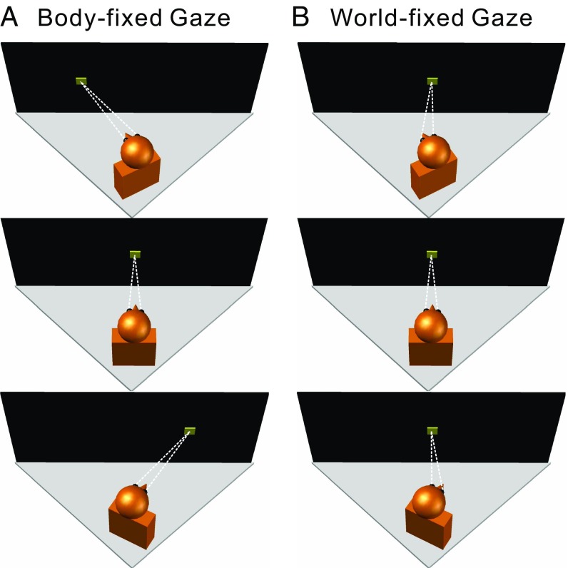
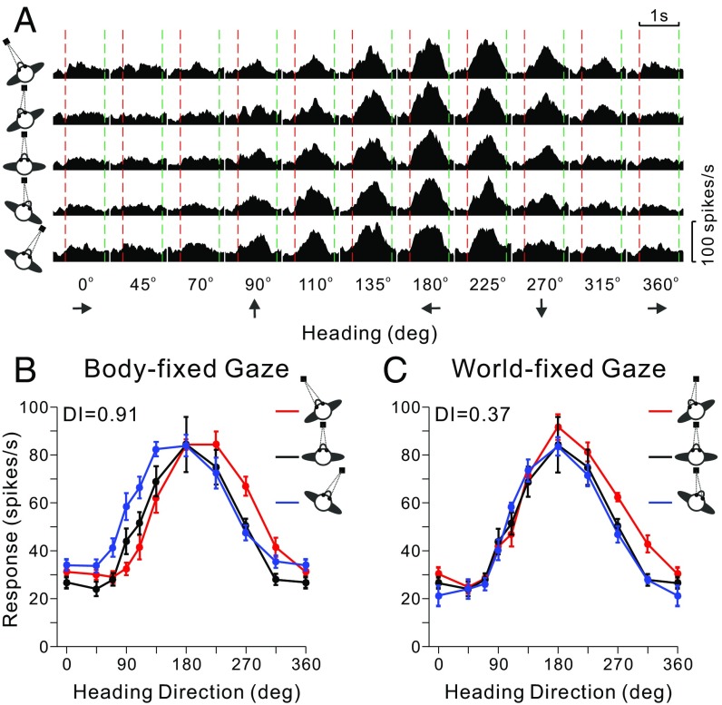
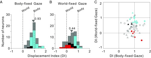
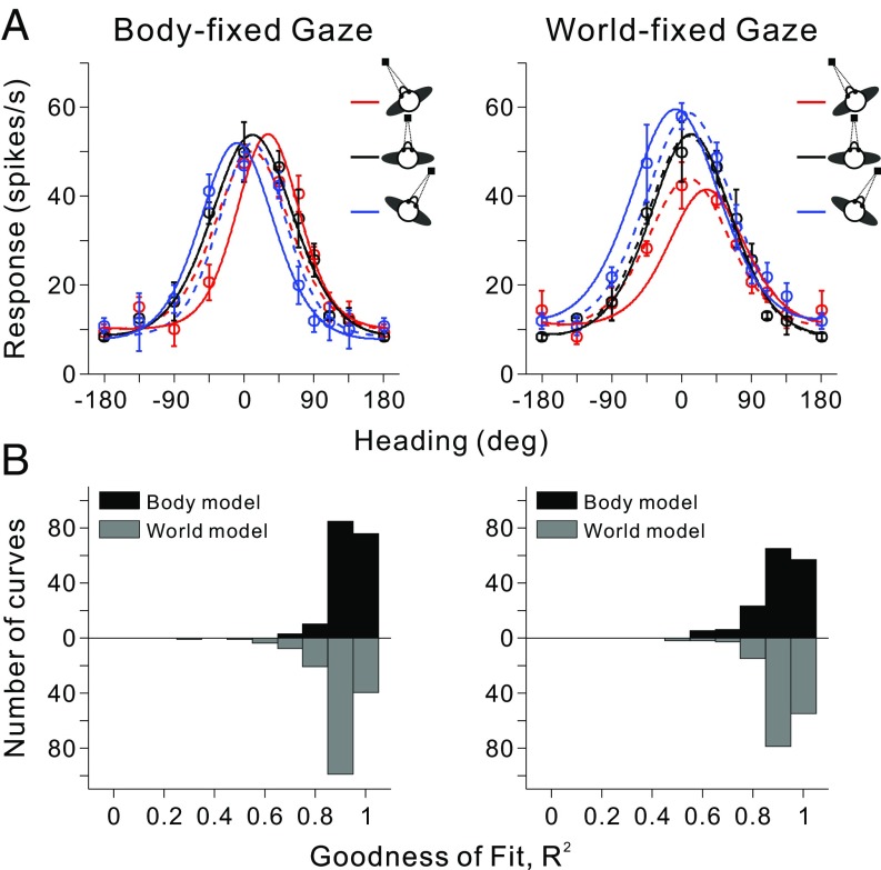
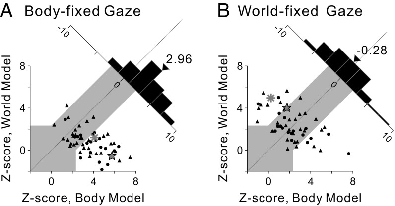
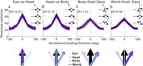
By systematically manipulating head position relative to the body and eye position relative to the head, previous studies have shown that vestibular tuning curves of neurons in the ventral intraparietal (VIP) area remain invariant when expressed in body-/world-centered coordinates. However, body orientation relative to the world was not manipulated; thus, an egocentric, body-centered representation could not be distinguished from an allocentric, world-centered reference frame. We manipulated the orientation of the body relative to the world such that we could distinguish whether vestibular heading signals in VIP are organized in body- or world-centered reference frames. We found a hybrid representation, depending on gaze direction. When gaze remained fixed relative to the body, the vestibular heading tuning of VIP neurons shifted systematically with body orientation, indicating an egocentric, body-centered reference frame. In contrast, when gaze remained fixed relative to the world, this representation changed to be intermediate between body- and world-centered. We conclude that the neural representation of heading in posterior parietal cortex is flexible, depending on gaze and possibly attentional demands.

Keywords: body/world-centered; egocentric/allocentric; reference frame; ventral intraparietal area; vestibular.


Conflict of interest statement

The authors declare no conflict of interest.

Figures

Fig. 1.
Fig. 1.
Experimental design. Schematic illustration of the various combinations of body/eye orientations relative to the motion platform (gray triangles) and display screen (black rectangle). The monkey’s head/body orientation was varied relative to the motion platform and screen (Top, 20° left; Middle, straight ahead, 0°; Bottom, 20° right) by using a yaw rotator that was mounted on top of the motion platform, below the monkey. The monkey’s head was fixed relative to the body, but the eye-in-head position could be manipulated by fixating one of three targets (yellow squares) on the screen. (A) In the body-fixed gaze condition, the monkey fixated a visual target that was at a fixed location (straight ahead) relative to the head/body, such that eye-in-head position was constant. (B) In the world-fixed gaze condition, the animal was required to maintain fixation on a world-fixed target, such that eye-in-head position covaried with head/body orientation.
Fig. 2.
Fig. 2.
Data from an example VIP neuron. (A) Response peristimulus time histograms are shown for the 10 headings (x axis) and each of the five combinations of [body-in-world, eye-in-head] positions: [−20°, 0°], [−20°, 20°], [0°, 0°], [20°, −20°], [20°, 0°] (rows, top to bottom). The red and green vertical dashed lines represent the start and end of platform motion (which followed a straight trajectory for each different heading with a Gaussian velocity profile). (B) Tuning curves for the body-fixed gaze condition. The three tuning curves show mean firing rate (±SEM) as a function of heading for the three combinations of [body-in-world, eye-in-head] position ([−20°, 0°], [0°, 0°], [20°, 0°]) that have constant eye-in-head position, as indicated by the red, black, and blue curves, respectively. (C) Tuning curves for the world-fixed gaze condition for the three combinations of [body-in-world, eye-in-head] position ([−20°, 20°], [0°, 0°], [20°, −20°]) that involve fixation on the same world-fixed target.
Fig. 3.
Fig. 3.
Summary of spatial reference frames for vestibular heading tuning in VIP, as quantified by the DI. (A and B) DI values of 1 and 0 indicate body- and world-centered tuning, respectively. Data are shown for both body-fixed gaze (A; n = 60) and world-fixed gaze (B; n = 61) conditions. Cyan and red bars in A and B represent neurons that are statistically classified as body- and world-centered, respectively (see Methods for details). Black bars represent neurons that are classified as intermediate, whereas gray bars represent neurons that are unclassified. Arrowheads indicate mean DI values for each distribution. Mean values of the DI distributions for monkeys F and X were not significantly different (body-fixed gaze: P = 0.06; world-fixed gaze: P = 0.28, t tests); thus, data for A and B were pooled across monkeys. (C) Scatter plot comparing DI values for the body- and world-fixed gaze conditions. Circles and triangles denote data from monkeys F and X, respectively. Colors indicate cells classified as body centered in both conditions (cyan), intermediate in both conditions (black), or body-centered in the body-fixed gaze condition but world-centered in the world-fixed gaze condition (red). Open symbols denote unclassified neurons (Table S1).
Fig. 4.
Fig. 4.
Model fits to vestibular heading tuning curves. (A) Heading tuning curves for an example VIP neuron (mean firing rate ± SEM) are shown for both the body-fixed (Left) and world-fixed (Right) gaze conditions. For each condition, the three tuning curves were fit simultaneously with body-centered (solid curves) and world-centered (dashed curves) models that are based on von Mises functions (see Methods for details). (B) Distributions of R2 values, which measure goodness-of-fit. For each condition, black bars represent fits with the body-centered model, whereas gray bars denote fits with the world-centered model (data for the world-centered model are plotted in the downward direction for ease of comparison). Note that only neurons with significant tuning for all three gaze positions have been included in this comparison (body-fixed gaze, n = 57; world-fixed gaze, n = 52). The median R2 values for monkeys F and X were not significantly different for either the body-fixed gaze condition (body-centered model, P = 0.57; world-centered model, P = 0.41; Wilcoxon rank sum test) or the world-fixed gaze condition (body-centered model, P = 0.67; world-centered model, P = 0.48); thus, data were pooled across animals in these distributions.
Fig. 5.
Fig. 5.
Model-based classification of spatial reference frames for vestibular heading tuning. Z-scored partial correlation coefficients between the data and the two models (body- and world-centered) are shown for the body-fixed gaze condition (A; n = 57) and the world-fixed gaze condition (B; n = 52). The gray region marks the boundaries of CIs that distinguish the models statistically. Circles and triangles denote data from monkeys F and X, respectively. The star and asterisk represent the example neurons of Figs. 2 and 4, respectively. Diagonal histograms represent distributions of differences between Z scores for the two models. Arrowheads indicate the mean values for each distribution. Median values of Z-scored partial correlation coefficients were not significantly different for monkeys F and X in either the body-fixed gaze condition (body-centered model, P = 0.11; world-centered model, P = 0.19; Wilcoxon rank-sum test) or in the world-fixed gaze condition (body-centered model, P = 0.86; world-centered model, P = 0.14); thus, data were pooled across animals.
Fig. 6.
Fig. 6.
Summary of vestibular heading representation in VIP. Average normalized heading tuning curves and DI values for data from Chen et al. (19) (A and B) and for the current data (C and D). Colored lines and error bands indicate the mean firing rate ± SEM. Vertical dotted lines show the direction around which tuning curves are aligned (see SI Methods for details). The colored arrows below each graph indicate the direction of reference frames in each condition. The direction of the world reference (thick black arrow) is the same in all schematics. In A (eye vs. head), eye position (cyan arrows) was varied, while head and body positions (magenta and gray arrows) were kept constant. In B (head vs. body), eye and head positions changed together, while body orientation was fixed. In C (body-fixed gaze), eye, head, and body all varied together. In D (world-fixed gaze), head and body changed, while eye position was fixed in the world.

Similar articles

Cited by

References

    1. Gu Y, Watkins PV, Angelaki DE, DeAngelis GC. Visual and nonvisual contributions to three-dimensional heading selectivity in the medial superior temporal area. J Neurosci. 2006;26:73–85. - PMC - PubMed
    1. Chen A, DeAngelis GC, Angelaki DE. Representation of vestibular and visual cues to self-motion in ventral intraparietal cortex. J Neurosci. 2011;31:12036–12052. - PMC - PubMed
    1. Chen A, DeAngelis GC, Angelaki DE. Macaque parieto-insular vestibular cortex: Responses to self-motion and optic flow. J Neurosci. 2010;30:3022–3042. - PMC - PubMed
    1. Gu Y, Cheng Z, Yang L, DeAngelis GC, Angelaki DE. Multisensory convergence of visual and vestibular heading cues in the pursuit area of the frontal eye field. Cereb Cortex. 2016;26:3785–3801. - PMC - PubMed
    1. Fan RH, Liu S, DeAngelis GC, Angelaki DE. Heading tuning in macaque area V6. J Neurosci. 2015;35:16303–16314. - PMC - PubMed

Publication types

LinkOut - more resources