. 2018 Apr 3;115(14):E3155-E3162.
doi: 10.1073/pnas.1704639115. Epub 2018 Mar 19.

Dimeric sorting code for concentrative cargo selection by the COPII coat

Affiliations

Dimeric sorting code for concentrative cargo selection by the COPII coat

Chao Nie et al. Proc Natl Acad Sci U S A. .

Abstract

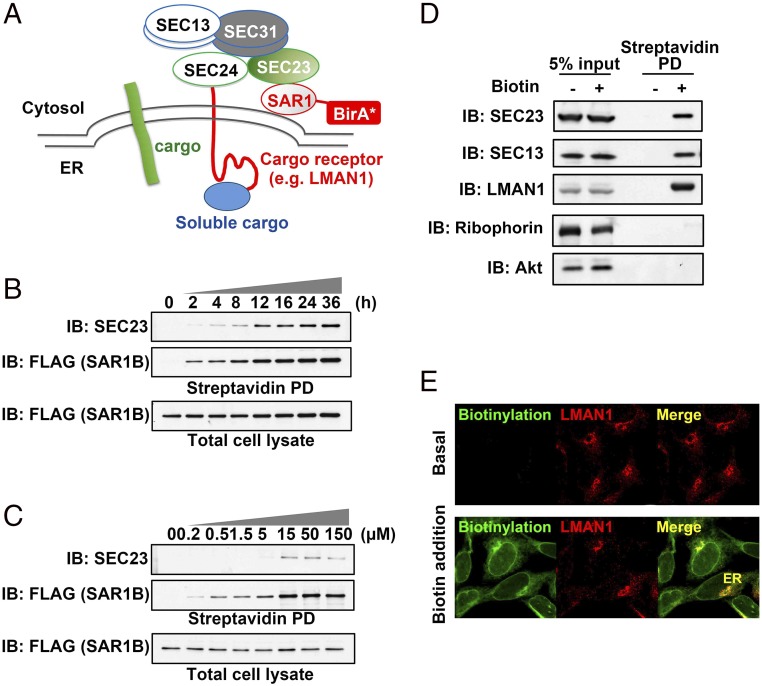
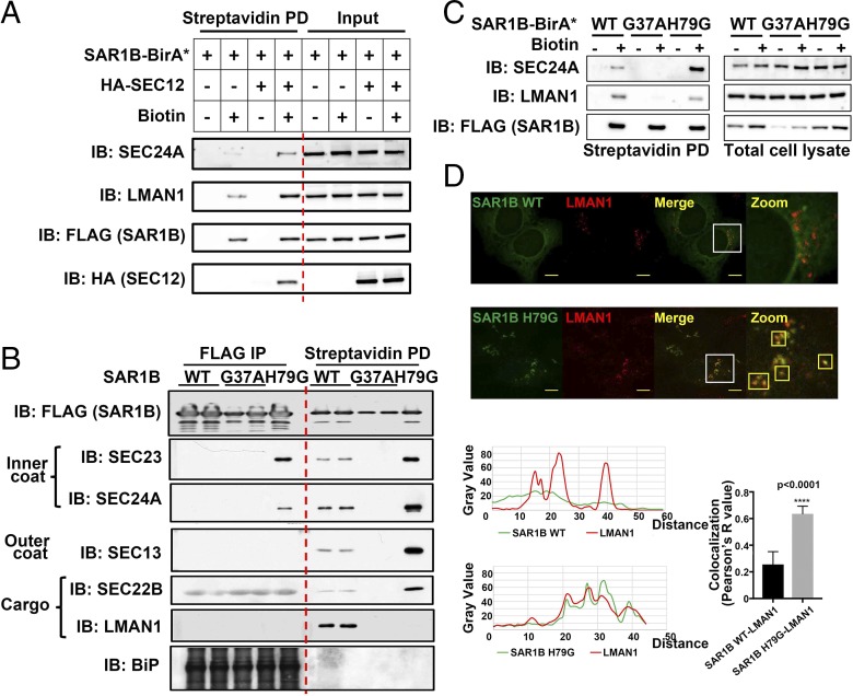
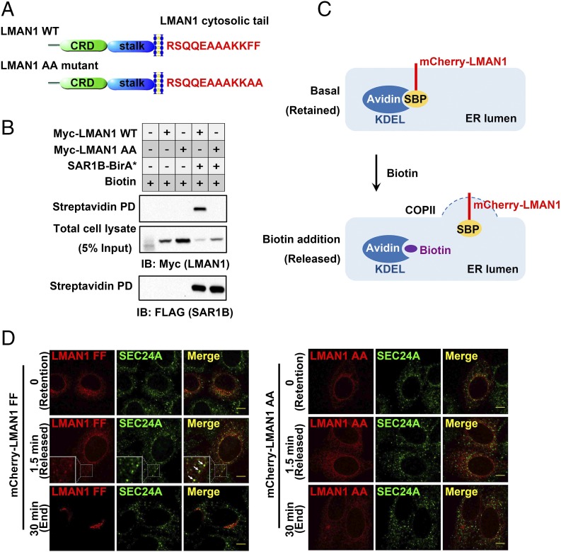
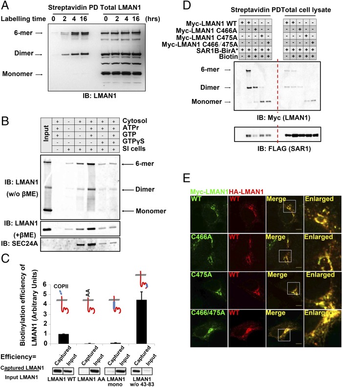
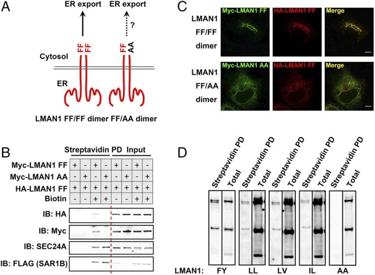
The flow of cargo vesicles along the secretory pathway requires concerted action among various regulators. The COPII complex, assembled by the activated SAR1 GTPases on the surface of the endoplasmic reticulum, orchestrates protein interactions to package cargos and generate transport vesicles en route to the Golgi. The dynamic nature of COPII, however, hinders analysis with conventional biochemical assays. Here we apply proximity-dependent biotinylation labeling to capture the dynamics of COPII transport in cells. When SAR1B was fused with a promiscuous biotin ligase, BirA*, the fusion protein SAR1B-BirA* biotinylates and thus enables the capture of COPII machinery and cargos in a GTP-dependent manner. Biochemical and pulse-chase imaging experiments demonstrate that the COPII coat undergoes a dynamic cycle of engagement-disengagement with the transmembrane cargo receptor LMAN1/ERGIC53. LMAN1 undergoes a process of concentrative sorting by the COPII coat, via a dimeric sorting code generated by oligomerization of the cargo receptor. Similar oligomerization events have been observed with other COPII sorting signals, suggesting that dimeric/multimeric sorting codes may serve as a general mechanism to generate selectivity of cargo sorting.

Keywords: COPII; LMAN1; cargo receptor; cargo sorting; proximity-dependent biotinylation.


Conflict of interest statement

The authors declare no conflict of interest.

Figures

Fig. 1.
Fig. 1.
Development of a proximity-dependent biotinylation assay for COPII-mediated cargo transport. (A) Overall scheme of proximity-dependent biotinylation of the COPII machinery using SAR1-BirA*. Activated SAR1 initiates the assembly of the COPII coat, which recruits cargos and/or additional regulatory factors (e.g., the cargo receptor LMAN1) to defined microdomains, allowing biotinylation by BirA* fused to SAR1. (B) Time course of biotinylation of the COPII subunit SEC23 by SAR1B-BirA*. 293A cells stably expressing SAR1B-BirA* (with a FLAG tag at the C terminus of BirA*) were treated with 15 µM biotin for different time points as indicated. After cell lysis, biotinylated proteins were isolated by streptavidin bead pull-down (PD) and subjected to SDS/PAGE followed by immunoblotting (IB) with anti-SEC23 (Top) or anti-FLAG (recognizing SAR1B; Middle). Immunoblotting for SAR1B-FLAG in total cell lysates is shown (Bottom). The anti-SEC23 antibody recognizes both SEC23A and SEC23B. (C) Biotin dose dependence of SEC23 labeling by SAR1B-BirA*. 293A cells stably expressing SAR1B-BirA* were treated for 4 h with different doses of biotin as indicated. After cell lysis, biotinylated proteins were isolated by streptavidin beads and subjected to SDS/PAGE followed by immunoblotting with the indicated antibodies. (D) Capture of COPII cargos by SAR1B-BirA* in vivo. 293A cells stably expressing SAR1-BirA* were treated with 15 µM biotin for 4 h. After cell lysis, biotinylated proteins were isolated by streptavidin beads and subjected to SDS/PAGE followed by immunoblotting with the indicated antibodies; “5% input” indicates total cell lysate equivalent to 5% of the material used for streptavidin pull-down in lanes 3 and 4 (“Streptavidin PD”). (E) Subcellular localization of biotinylated proteins. 293A cells stably expressing SAR1B-BirA* were treated with 15 µM biotin for 4 h. After cell fixation, biotinylated proteins were visualized by immunostaining and confocal microscopy with an anti-LMAN1 antibody or Alexa Fluor-conjugated streptavidin.
Fig. 2.
Fig. 2.
Assembly–disassembly cycle of COPII revealed by the proximity assay. (A) The SAR1 activator SEC12 promotes labeling of COPII subunits and cargos. 293A cells transfected with the indicated DNA constructs were treated with 15 µM biotin for 4 h. After cell lysis, biotinylated proteins were isolated by streptavidin beads and subjected to SDS/PAGE followed by immunoblotting with the indicated antibodies. (B) GTP-dependent cycling of the COPII coats revealed by coimmunoprecipitation (Left) and biotinylation (Right). 293A cells stably expressing the indicated SAR1B-BirA*-FLAG constructs were treated with 15 µM biotin for 4 h. After cell lysis, proteins were isolated by mouse anti-FLAG beads (FLAG IP) or streptavidin beads (Streptavidin PD) and subjected to SDS/PAGE followed by immunoblotting with the indicated antibodies. Mouse antibodies against BiP yielded strong nonspecific signal with the mouse anti-FLAG IgG used for IP. (C) GTP-locked SAR1B (H79G) decreases LMAN1 biotinylation. 293A cells transfected with the indicated DNA constructs were treated with 15 µM biotin for 4 h. After cell lysis, biotinylated proteins were isolated by streptavidin beads and subjected to SDS/PAGE followed by immunoblotting with the indicated antibodies. (D) GTP-locked SAR1B (H79G) traps LMAN1 on the ER. 293A cells stably expressing SAR1-BirA* or the H79G mutant were fixed and visualized by immunostaining and confocal microscopy with the indicated antibodies. Smaller boxes outlined in yellow indicate colocalization of LMAN1 and SAR1B. (Scale bars, 8 μm.) (D, Bottom) Quantification of the colocalization of green (SAR1B) and red (LMAN1) fluorophores. A total of 10 cells was analyzed for SAR1B H79G and for SAR1B WT. Error bars represent SEM.
Fig. 3.
Fig. 3.
Concentrative sorting of LMAN1 by COPII coats. (A) Schematics of LMAN1. The luminal domains and amino acid sequences of the cytosolic tails in wild-type LMAN1 and the AA mutant are shown. CRD, carbohydrate-binding domain. The stalk domain mediates LMAN1 oligomerization (51). (B) Enrichment of wild-type LMAN1, but not the LMAN1-AA mutant, by COPII revealed by SAR1B-BirA*. 293A cells stably expressing SAR1B-BirA* were transfected with the indicated LMAN1 constructs and treated with 15 µM biotin for 24 h. After cell lysis, biotinylated proteins were isolated by streptavidin beads and subjected to SDS/PAGE followed by immunoblotting with the indicated antibodies. (C) Schematics of the pulse–chase experiment using the RUSH imaging strategy. In the basal state (no biotin), SBP-mCherry-LMAN1 is retained in the ER by streptavidin-KDEL. Addition of biotin releases SBP-mCherry-LMAN1 from streptavidin-KDEL, allowing LMAN1 to be exported from the ER. (D) Wild-type LMAN1, but not the LMAN1-AA mutant, is enriched on COPII-coated puncta before ER export. HeLa cells expressing the indicated SBP-mCherry-LMAN1 constructs and streptavidin-KDEL were fixed at different time points following biotin treatment and subjected to immunostaining with an anti-SEC24A antibody, followed by confocal microscopy. Arrows indicate the colocalization of LMAN1 and SEC24A on the ER surface. (Scale bars, 8 µm.)
Fig. 4.
Fig. 4.
LMAN1 dimers constitute the minimal unit for export from the ER. (A) ER export of LMAN1 dimers and oligomers revealed by the proximity assay. 293A cells stably expressing SAR1B-BirA* were treated with 15 µM biotin for the indicated time points. After cell lysis, biotinylated proteins were isolated by streptavidin beads and subjected to SDS/PAGE without reducing agents followed by immunoblotting with an anti-LMAN1 antibody. (B) Cell-free “budding” assay to reconstitute ER export of LMAN1 dimers/oligomers. Semiintact (SI) cells were mixed with cytosol and the indicated reagents to catalyze in vitro vesicle formation from the ER. The isolated vesicles were subjected to immunoblotting without reducing agent (Top) or with reducing agent (Middle and Bottom) with the indicated antibodies. No dimers or hexamers of LMAN1 were observed in the reducing condition. ATPr, ATP regeneration system. GTPγS is the nonhydrolyzable analog of GTP, and blocks COPII budding from the ER. (C) Biotinylation efficiency of LMAN1 mutants in the proximity assay. 293A cells stably expressing SAR1B-BirA* were transfected with the indicated Myc-LMAN1 mutants and treated with 15 µM biotin for 4 h. After cell lysis, biotinylated proteins were isolated by streptavidin beads and subjected to SDS/PAGE followed by immunoblotting with an anti-Myc antibody. (C, Top) LMAN1 is depicted in red, with blue bars representing deleted regions within the LMAN1 protein. Error bars represent SEM. (D) Dimeric LMAN1 is biotinylated by SAR1B-BirA*. 293A cells stably expressing SAR1B-BirA* were transfected with the indicated Myc-LMAN1 cysteine-to-alanine mutants and treated with 15 µM biotin for 4 h. After cell lysis, biotinylated proteins were isolated by streptavidin beads and subjected to native SDS/PAGE followed by immunoblotting with an anti-Myc antibody. Different oligomerization states of LMAN1 are depicted (Left). (E) Dimeric LMAN1 is targeted to the ERGICs. Cos-1 cells expressing the indicated LMAN1 constructs were fixed and subjected to immunostaining with the indicated antibodies, followed by confocal microscopy. (Scale bars, 8 µm.)
Fig. 5.
Fig. 5.
Dimeric sorting motifs mediate ER export via the COPII coat. (A) Schematics of a homodimeric FF–FF motif or a heterodimeric FF–AA motif. (B) LMAN1-AA inhibits ER export of the wild-type LMAN1 revealed by the proximity assay. 293A cells stably expressing SAR1B-BirA* were transfected with the indicated LMAN1 mutants and treated with 15 µM biotin for 4 h. After cell lysis, biotinylated proteins were isolated by streptavidin beads and subjected to SDS/PAGE followed by immunoblotting with the indicated antibodies. (C) LMAN1-AA traps wild-type LMAN1 in the ER. HeLa cells expressing the indicated LMAN1 constructs were fixed and subjected to immunostaining with the indicated antibodies, followed by confocal microscopy. (Scale bars, 8 µm.) (D) Dimeric FY, LL, LV, and IL sorting motifs in ER export. 293A cells stably expressing SAR1-BirA* were transfected with Myc-LMAN1 mutants with the indicated sorting motifs and treated with 15 µM biotin for 4 h. After cell lysis, biotinylated proteins were isolated by streptavidin beads and subjected to SDS/PAGE followed by immunoblotting with an anti-Myc antibody.

Similar articles

Cited by

References

    1. Bonifacino JS, Glick BS. The mechanisms of vesicle budding and fusion. Cell. 2004;116:153–166. - PubMed
    1. Palade G. Intracellular aspects of the process of protein synthesis. Science. 1975;189:347–358. - PubMed
    1. Barlowe C, Helenius A. Cargo capture and bulk flow in the early secretory pathway. Annu Rev Cell Dev Biol. 2016;32:197–222. - PubMed
    1. Cai H, Reinisch K, Ferro-Novick S. Coats, tethers, Rabs, and SNAREs work together to mediate the intracellular destination of a transport vesicle. Dev Cell. 2007;12:671–682. - PubMed
    1. Brandizzi F, Barlowe C. Organization of the ER-Golgi interface for membrane traffic control. Nat Rev Mol Cell Biol. 2013;14:382–392. - PMC - PubMed

Publication types

LinkOut - more resources