. 2018 May;177(1):381-397.
doi: 10.1104/pp.17.01714. Epub 2018 Mar 19.

Ribosomal RNA Biogenesis and Its Response to Chilling Stress in Oryza sativa

Affiliations

Ribosomal RNA Biogenesis and Its Response to Chilling Stress in Oryza sativa

Runlai Hang et al. Plant Physiol. 2018 May.

Abstract

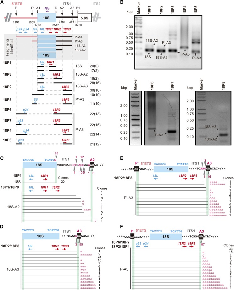
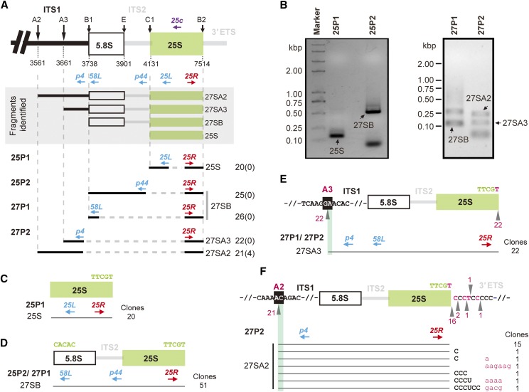
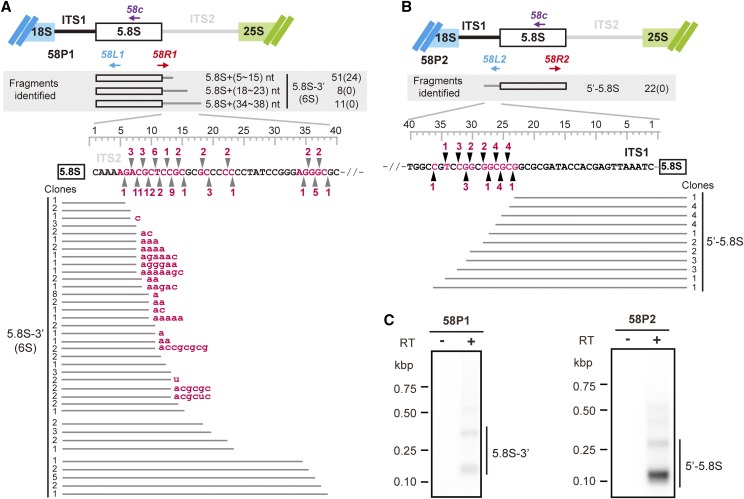
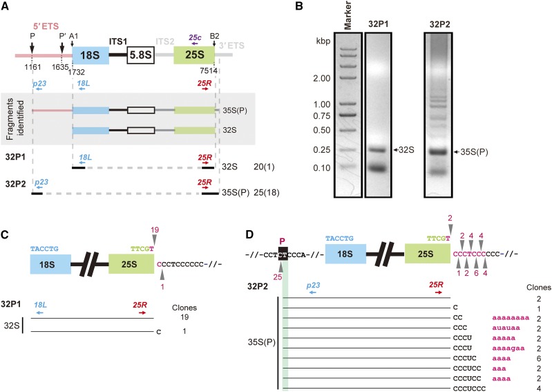
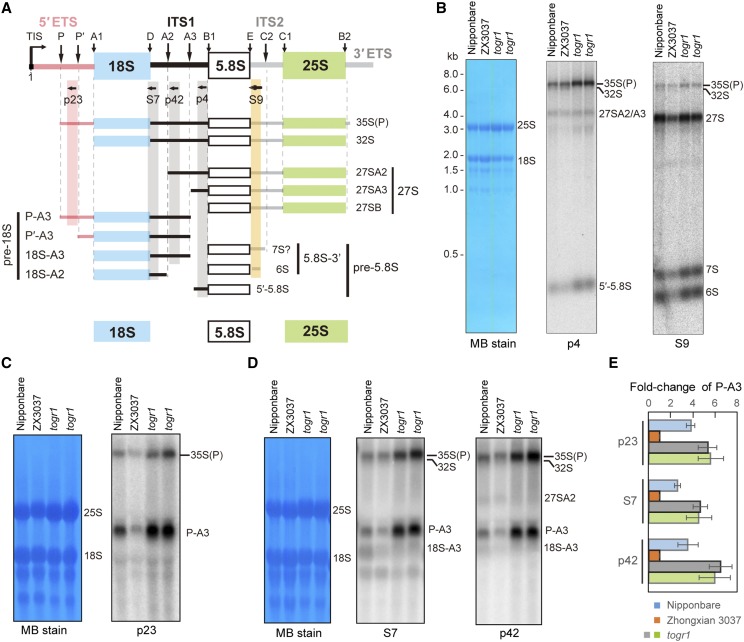
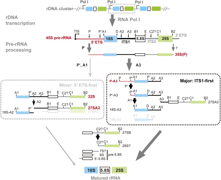
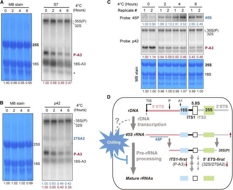
Ribosome biogenesis is crucial for plant growth and environmental acclimation. Processing of ribosomal RNAs (rRNAs) is an essential step in ribosome biogenesis and begins with transcription of the rDNA. The resulting precursor-rRNA (pre-rRNA) transcript undergoes systematic processing, where multiple endonucleolytic and exonucleolytic cleavages remove the external and internal transcribed spacers (ETS and ITS). The processing sites and pathways for pre-rRNA processing have been deciphered in Saccharomyces cerevisiae and, to some extent, in Xenopus laevis, mammalian cells, and Arabidopsis (Arabidopsis thaliana). However, the processing sites and pathways remain largely unknown in crops, particularly in monocots such as rice (Oryza sativa), one of the most important food resources in the world. Here, we identified the rRNA precursors produced during rRNA biogenesis and the critical endonucleolytic cleavage sites in the transcribed spacer regions of pre-rRNAs in rice. We further found that two pre-rRNA processing pathways, distinguished by the order of 5' ETS removal and ITS1 cleavage, coexist in vivo. Moreover, exposing rice to chilling stress resulted in the inhibition of rRNA biogenesis mainly at the pre-rRNA processing level, suggesting that these energy-intensive processes may be reduced to increase acclimation and survival at lower temperatures. Overall, our study identified the pre-rRNA processing pathway in rice and showed that ribosome biogenesis is quickly inhibited by low temperatures, which may shed light on the link between ribosome biogenesis and environmental acclimation in crop plants.


Figures

Figure 1.
Figure 1.
Mapping of the 5′ and 3′ extremities of the pre-18S rRNAs. A, Structure of pre-18S rRNA intermediates identified by a set of primer combinations (in shaded box). Forward and reverse PCR primers for cDNA amplification are marked in red and blue, respectively. For each fragment, the number of clones obtained is indicated on the right. The number of clones with additional sequences, such as polyadenylation at the 3′ end, is marked in parentheses. Eight pairs of primers were used: 18P1 (18L/18R1), 18P2 (18L/18R3), 18P3 (p23/18R3), 18P4 (p24/18R3), 18P5 (S5/18R3), 18P6 (p24/18R2), 18P7 (p23/18R2), and 18P8 (18L/18R2). B, Pre-18S rRNA intermediates were determined in gel by cRT-PCR with primers 18P1 to 18P8. C to F, DNA sequencing of 18S and its major precursors identified: 18S-A2 (C), 18S-A3 (D), P′-A3 (E), and P-A3 (F). The 18S rRNAs identified by primers 18P1 were validated by sequencing of 20 independent clones. The 18S-A2 intermediates identified by primers 18P1 and 18P8 were validated by sequencing of 33 independent clones (C). The 18S-A3 intermediates identified by primers 18P2 and 18P8 were validated by sequencing of 58 independent clones (D). The P′-A3 intermediates identified by primers 18P2 and 18P8 were validated by sequencing of 21 independent clones (E). The P-A3 intermediates identified by primers 18P6, 18P7, 18P3, and 18P4 were validated by sequencing of 87 independent clones (F). The ITS1 locus matched by the 3′ ends of these clones are indicated by black triangles as well as the number of clones. Additional sequences in the 3′ extremities of these clones are marked in red lowercase letters. The numbers of identical clones are indicated to the right of each fragment.
Figure 2.
Figure 2.
Mapping of the 5′ and 3′ extremities of the pre-25S rRNAs. A, Structure of pre-25S intermediates identified by a set of primers (in shaded box). Forward and reverse PCR primers for cDNA amplification are marked in red and blue, respectively. Four pairs of primers were used for pre-25S rRNAs: 25P1 (25L/25R), 25P2 (p44/25R), 27P1 (58L/25R), and 27P2 (p4/25R). For each fragment, the number of clones obtained is indicated on the right. The number of clones containing additional sequences at the 3′ extremities is marked in parentheses. B, Pre-25S rRNA intermediates were determined in gel by cRT-PCR with primers 25P1, 25P2, 27P1, and 27P2. C to F, The DNA sequencing results for 25S (C) and its major precursors identified: 27SB (D), 27SA3 (E), and 27SA2 (F). The 25S rRNA identified by primers 25P1 were validated by sequencing of 20 independent clones (C). The 27SB intermediates identified by primers 25P2 and 27P1 were validated by sequencing of 51 independent clones (D). The 27SA3 intermediates identified by primers 27P1 and 27P2 were validated by sequencing of 22 independent clones (E). The 27SA2 intermediates identified by primers 27P2 were validated by sequencing of 21 independent clones (F). The ITS1 and ITS2 locus matched by the 5′ and 3′ ends of these DNA sequences, respectively, are indicated by black triangles as well as the number of clones. Additional sequences in the 3′ extremities of these clones are marked in red lowercase letters. The number of identical clones is indicated to the right of each fragment.
Figure 3.
Figure 3.
Mapping of the 5′ and 3′ extremities of the pre-5.8S rRNAs. A and B, Structure of 3′-5.8S identified by 58P1 (58L1/58R1; A) and 5′-5.8S by 58P2 (58L2/58R2; B), respectively. Forward and reverse PCR primers for cDNA amplification are marked in red and blue, respectively. For each fragment, the number of clones obtained is indicated on the right. The number of clones containing additional sequences at the 3′ extremities are marked in parentheses (in the shaded box). The 5.8S-3′ intermediates were validated by 70 independent clones (A). The 5′-5.8S intermediates were validated by 22 independent clones (B). The ITS1 and ITS2 locus matched by the 5′ and 3′ ends of these DNA sequences, respectively, are indicated by black triangles as well as the number of clones. Additional sequences in the 3′ extremities of these clones are marked in red lowercase letters. The number of identical clones are indicated to the left (A) and right (B) of each fragment, respectively. C, Pre-5.8S rRNA intermediates were determined in gel by cRT-PCR with primers 58P1 and 58P2.
Figure 4.
Figure 4.
Mapping of the 5′ and 3′ extremities of the 35S(P) and 32S transcripts. A, Structure of early pre-rRNA intermediates identified (in shaded box) by two pairs of primers: 32P1 and 32P2. Forward and reverse PCR primers for cDNA amplification are marked in red and blue, respectively. For each fragment, the number of clones obtained is indicated on the right. The number of clones with additional sequences at the 3′ end is marked in parentheses. B, The 32S and 35S(P) pre-rRNAs were determined in gel by cRT-PCR with primers 32P1 (18L/25R) and 32P2 (p23/25R). C and D, DNA sequencing results for 32S (C) and 35S(P) precursors (D). The 32S pre-rRNAs were validated by sequencing of 20 independent clones (D). The 35S(P) pre-rRNAs were validated by sequencing of 25 independent clones (D). The ITS1 and ITS2 locus matched by the 5′ and 3′ ends of these DNA sequences, respectively, are indicated by black triangles and the number of clones. Additional sequences in the 3′ extremities of these clones are marked in red lowercase letters. The number of identical clones is indicated to the right of each fragment.
Figure 5.
Figure 5.
Northern blots to detect pre-rRNA processing in rice. A, Pre-rRNA processing intermediates detected by northern blots with specific probes, which are indicated by horizontal arrows. Black vertical arrows above the diagram indicate endonucleolytic cleavage sites relevant to this study. Different rRNA precursors are marked. P-A3, P′-A3, 18S-A3, and 18S-A2 belong to the pre-18S rRNAs. 27SA2, 27SA3, and 27SB belong to the 27S rRNA, the common precursor of 5.8S and 25S rRNAs. The 3′-5.8S (7S and 6S) and 5′-5.8S are pre-5.8S rRNAs. The 7S rRNA marked with “?” was detected by probe S9 (Fig. 5B), but its definite 3′ extremities are still unclear (A). The 35S(P) and 27SA2 could be specifically detected by probes p23 and p42, respectively. Both probes S7 and p42 detect 35S(P), 32S, P-A3, and 18S-A3. Although 18S-A2 could be detected by S7, its low abundance in wild-type rice makes it harder to distinguish from 18S-A3 by northern-blot assay. B, Northern blots to determine pre-rRNA processing in pre-60S LSU in Nipponbare (lane 1), Zhongxian3037 (ZX3037, lane 2), and togr1 mutants (lanes 3 and 4). The togr1 mutant is a positive control that accumulates the 35S pre-rRNA and P-A3 intermediates, when compared with its wild type, Zhongxian3037 (Wang et al., 2016). Probes p4 and S9 were used. Methylene blue staining (MB stain) of the membrane is shown as the loading control. C to E, Northern blots to determine pre-rRNA processing in pre-40S SSU by probes p23 (C), S7, and p42 (D) in rice. The S7 and p42 blots share the same loading control (D). The quantitation of P-A3 in Nipponbare (lane 1), Zhongxian3037 (lane 2), and togr1 (lanes 3 and 4) were performed with three biological replicates (E). Matured rRNAs stained with MB serve as the loading control. The relative intensities for P-A3 intermediate in each lane are normalized to Zhongxian3037. Error bars represent sd. Data are given as means and sd of three independent biological replicates.
Figure 6.
Figure 6.
Model of rRNA biogenesis in rice. Primary transcripts generated by RNA Polymerase I are first processed at P in the 5′ ETS and at an unknown site in the 3′ ETS to generate 35S(P), which undergoes further pre-rRNA processing by alternative pathways distinguished by the order of ITS1 splitting and 5′ ETS removal, to generate mature 18S, 5.8S, and 25S rRNAs. In the major ITS1-first pathway, the 35SP transcript is split at ITS1 endonucleolytic site A3 into P-A3 and 27SA3 precursors. In the minor 5′ ETS-first pathway, the removal of the 5′ ETS in the 35S(P) transcript occurs first to generate the 32S intermediate before its split at the ITS1 cleavage site A2. Both endo- and exonucleolytic processing occur sequential and coordinately in this progress. Precursors with partial transparency indicate putative intermediates in these pathways.
Figure 7.
Figure 7.
Chilling stress inhibits rRNA biogenesis mainly at pre-rRNAs processing levels. A and B, Northern blots to detect pre-rRNA processing in Nipponbare (japonica) rice under 4°C treatment for 0, 2, 4, and 6 h, with probes S7 (A) and p42 (B). Matured rRNAs stained with MB serve as the loading control. The numbers below each lane represent the intensity ratio of each signal relative to the 0 h sample. The relative intensities for 25S rRNA, P-A3, and 27SA2 intermediates are marked in black, red, and blue, respectively. The asterisk detected by probe S7 represents the mature 16S rRNAs. Three biological replicates were performed and a representative result is shown here. C, Northern blots to detect the 45S rRNA transcript by probe 45P in Nipponbare under 4°C treatment for 0, 2, 4, and 6 h. Both blots of 45P and p42 came from the same membrane. Matured rRNAs stained with MB serve as the loading control. The numbers below each lane represent the intensity ratio of each signal relative to the 0 h sample. The relative intensities for 25S rRNA, 45S transcripts, and P-A3 intermediates are marked in black, blue, and red, respectively. RNA samples from two biological replicates were loaded and detected in parallel. D, Simplified model that the inhibition of rRNA biogenesis in rice by chilling stress predominantly occurs at posttranscriptional level. The 45S rRNA, transcribed by RNA Pol I from rDNAs, undergoes pre-rRNA processing to release mature rRNAs. The steady level of 45S rRNA in vivo is the net product of rDNA transcription and subsequent pre-rRNA processing. Chilling stress inhibits pre-rRNA processing, shown by the time-course reduction of P-A3 and 27SA2 in both ITS1-first and 5′ ETS-first processing pathways, respectively (A and B). Although it remains unknown whether and how chilling treatment affect rDNA transcription, the increased 45S rRNA (C) could mainly originate from reduced pre-rRNA processing under chilling stress. The long probe 45P could distinguish the 45S rRNA from its product 35S(P).

Similar articles

Cited by

References

    1. Abbasi N, Kim HB, Park NI, Kim HS, Kim YK, Park YI, Choi SB (2010) APUM23, a nucleolar Puf domain protein, is involved in pre-ribosomal RNA processing and normal growth patterning in Arabidopsis. Plant J 64: 960–976 - PubMed
    1. Al Refaii A, Alix JH (2009) Ribosome biogenesis is temperature-dependent and delayed in Escherichia coli lacking the chaperones DnaK or DnaJ. Mol Microbiol 71: 748–762 - PubMed
    1. Anger AM, Armache JP, Berninghausen O, Habeck M, Subklewe M, Wilson DN, Beckmann R (2013) Structures of the human and Drosophila 80S ribosome. Nature 497: 80–85 - PubMed
    1. Bai D, Zhang J, Li T, Hang R, Liu Y, Tian Y, Huang D, Qu L, Cao X, Ji J, Zheng X (2016) The ATPase hCINAP regulates 18S rRNA processing and is essential for embryogenesis and tumour growth. Nat Commun 7: 12310. - PMC - PubMed
    1. Bailey-Serres J, Sorenson R, Juntawong P (2009) Getting the message across: cytoplasmic ribonucleoprotein complexes. Trends Plant Sci 14: 443–453 - PubMed

Publication types

MeSH terms

LinkOut - more resources