. 2018 May;177(1):241-254.
doi: 10.1104/pp.18.00010. Epub 2018 Mar 19.

Protein Storage Vacuoles Originate from Remodeled Preexisting Vacuoles in Arabidopsis thaliana

Affiliations

Protein Storage Vacuoles Originate from Remodeled Preexisting Vacuoles in Arabidopsis thaliana

Mistianne Feeney et al. Plant Physiol. 2018 May.

Abstract

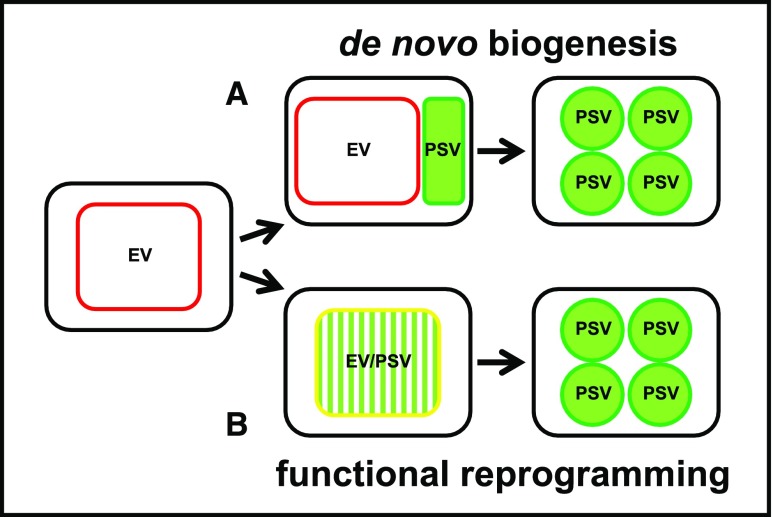
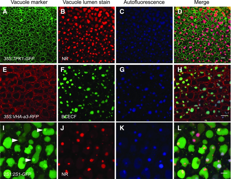
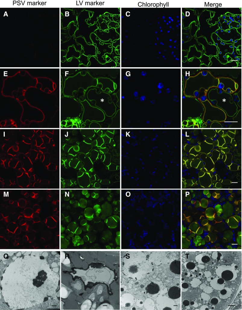
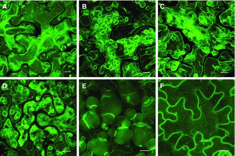
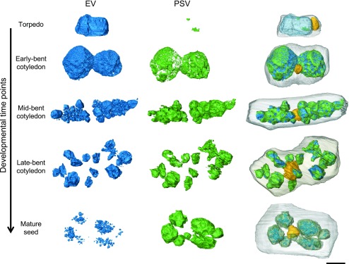
Protein storage vacuoles (PSV) are the main repository of protein in dicotyledonous seeds, but little is known about the origins of these transient organelles. PSV are hypothesized to either arise de novo or originate from the preexisting embryonic vacuole (EV) during seed maturation. Here, we tested these hypotheses by studying PSV formation in Arabidopsis (Arabidopsis thaliana) embryos at different stages of seed maturation and recapitulated this process in Arabidopsis leaves reprogrammed to an embryogenic fate by inducing expression of the LEAFY COTYLEDON2 transcription factor. Confocal and immunoelectron microscopy indicated that both storage proteins and tonoplast proteins typical of PSV were delivered to the preexisting EV in embryos or to the lytic vacuole in reprogrammed leaf cells. In addition, sectioning through embryos at several developmental stages using serial block face scanning electron microscopy revealed the 3D architecture of forming PSV. Our results indicate that the preexisting EV is reprogrammed to become a PSV in Arabidopsis.


Figures

Figure 1.
Figure 1.
Hypotheses for PSV formation tested in this work. A, PSV form de novo, briefly coexisting with the EV to eventually become the dominant structure in mature seeds. During the transition, PSV-specific tonoplast markers (green outline) appear alongside the EV tonoplast markers (red outline). B, PSV arise through reprogramming of the EV by the accumulation of seed storage proteins in the EV lumen (green pattern fill). PSV and EV tonoplast markers coexist (yellow outline) while the EV is converted to a PSV.
Figure 2.
Figure 2.
Stages assigned to Arabidopsis embryo development and an overview of PSV formation in a representative cell from selected stages. A, Images of heart, torpedo, walking stick, and bent cotyledon embryos dissected from Arabidopsis ovules. The mature embryo was dissected from a dry seed. Bar = 100 µm. B, PSV arise during the maturation phase of embryonic development. Images shows single micrographs from SBF-SEM stacks of embryos at different developmental stages. Deposits of electron-opaque material (arrows) are first observed in the vacuole lumen and along the tonoplast of the EV in early-bent cotyledon embryos. The deposits accumulate and eventually fill the vacuolar lumen in mature seed. Asterisks indicate oil bodies. Bar at left for high-magnification images = 1 µm; bar at right for inset overview images = 100 µm.
Figure 3.
Figure 3.
PSV tonoplast markers appear on the preexisting EV tonoplast in bent cotyledon embryos. Embryos constitutively expressing the EV tonoplast marker 35S:TPK1-GFP (green) and the PSV tonoplast markers (red) TIP3;1:TIP3;1-YFP (top and middle rows) or TIP3;2:TIP3;2-mCherry (bottom row) are shown. Chlorophyll autofluorescence is shown in blue. A to D, In early-bent cotyledon embryos, the EV is labeled with TPK1-GFP before PSV markers are expressed. E to L, In late-bent cotyledon embryos, PSV tonoplast markers colocalize with the EV tonoplast marker. Bars = 5 μm.
Figure 4.
Figure 4.
2S1 albumin seed storage proteins accumulate initially in punctate cytosolic structures and ultimately as deposits inside the lumina of forming PSV in bent cotyledon embryos. 2S1-GFP (green) labels small punctate structures (arrows) that accumulate in the EV/PSV lumen (asterisk). The PSV tonoplast marker TIP3;2-mCherry (red) is localized to the ER, tonoplast, and plasma membrane (open arrowheads). Chlorophyll is shown in blue. A to D, 2S1-GFP signal is first observed as small punctate structures in the cytoplasm and accumulates in PSV lumina whose tonoplasts are labeled with TIP3;2-mCherry. E to H, In late-bent cotyledon embryos, the 2S1-GFP signal is observed only in vacuole lumina. Subregions of more intense 2S1-GFP fluorescence are visible in PSV lumina (closed arrowheads). Bars = 5 µm.
Figure 5.
Figure 5.
Forming PSV are identified by the acidotropic stains NR and BCECF-AM and PSV luminal autofluorescence in bent cotyledon embryos. A to H, NR and BCECF-AM stain vacuoles labeled with the tonoplast markers TPK1-GFP (A) and VHA-a3-RFP (E), respectively. Vacuole lumen autofluorescence (blue) colocalizes with the stains (D and H). I to L, Embryos accumulating 2S1 albumin-GFP were stained with NR. The 2S1-GFP signal fills vacuole lumina, and areas of more intense GFP fluorescence are observed (arrowheads in I). NR stains distinct subregions of the vacuole lumina (J). These NR-stained subregions colocalize with PSV lumen autofluorescence (K) and with areas of intense 2S1-GFP fluorescence (L). Bars = 10 μm (A–H) and 5 μm (I–l).
Figure 6.
Figure 6.
Immunogold labeling reveals the localization of PSV tonoplast aquaporin TIP3;1 and the 2S albumin and 12S globulin seed storage proteins to the EV in bent cotyledon embryo cells. A and B, Anti-TIP3;1 antibody labels the tonoplast of transitioning vacuoles. C and D, Anti-12S globulin antibody labels electron-opaque material accumulating along the luminal side of the tonoplast (C) and the entire PSV lumen in late-bent cotyledon embryos (D). E and F, Anti-2S antibody labels electron-opaque material accumulating along the luminal side of the tonoplast (E) as well as electron-opaque material in the vacuole lumen (F). OB, Oil bodies. Bar = 500 nm.
Figure 7.
Figure 7.
PSV form through remodeling of the LV in Arabidopsis leaf cells reprogrammed by LEC2. A to P, Representative images of transitioning vacuoles in LEC2-induced leaf cells at 14 d (A–D), 17 d (E–H), and 20 d (I–P) with DEX. The TIP3;1-YFP PSV tonoplast marker (red) accumulates on the preexisting LV (E, I, and M) in Arabidopsis lines constitutively expressing the tonoplast marker TPK1-GFP (green). Chlorophyll autofluorescence is shown in blue. Asterisks show asynchronous remodeling of the tonoplast in neighboring cells. Bars = 10 μm. Q to T, Electron microscopy of leaf cells 14 d after LEC2 induction with DEX. Electron-opaque PSV material (black) accumulates along the luminal side of the tonoplast and disperses in the vacuole lumen. Bars = 500 nm (Q–S) and 2 μm (T).
Figure 8.
Figure 8.
The tonoplast undergoes extensive remodeling during the LV-to-PSV transition in LEC2-induced leaf cells. Representative images show the progression of tonoplast remodeling during the LV-to-PSV transition at 14 d (A), 17 d (B and C), and 20 d (D and E) with DEX or at 14 d without DEX (F). Arabidopsis 35S:LEC2-GR lines harboring 35S:TPK1-GFP (green) were imaged. Images are maximum intensity projections of Z-stacks taken through leaf epidermal cells. Bars = 10 µm.
Figure 9.
Figure 9.
Remodeling of the EV to PSV during embryo maturation. The lumen of the EV (blue), electron-opaque PSV luminal material (green), nucleus (yellow), and plasma membrane (gray) were rendered from SBF-SEM image stacks in cotyledon cells of embryos in the torpedo, early-, mid-, and late-bent cotyledon, or mature embryo stages. As electron-opaque material initially accumulates along the periphery of the EV lumen, the vacuole separates into several smaller vacuoles, which are eventually filled with electron-opaque PSV luminal material aside from small translucent areas. The nucleus changes position from the cell cortex to the center of the cell. Bar = 5 μm.

Comment in

  • Turnover of Tonoplast Proteins.
    Ivanov R, Robinson DG. Ivanov R, et al. Plant Physiol. 2018 May;177(1):10-11. doi: 10.1104/pp.18.00322. Plant Physiol. 2018. PMID: 29720533 Free PMC article. No abstract available.

Similar articles

Cited by

References

    1. Bagga S, Sutton D, Kemp JD, Sengupta-Gopalan C (1992) Constitutive expression of the β-phaseolin gene in different tissues of transgenic alfalfa does not ensure phaseolin accumulation in non-seed tissue. Plant Mol Biol 19: 951–958 - PubMed
    1. Baud S, Kelemen Z, Thévenin J, Boulard C, Blanchet S, To A, Payre M, Berger N, Effroy-Cuzzi D, Franco-Zorrilla JM, et al. (2016) Deciphering the molecular mechanisms underpinning the transcriptional control of gene expression by master transcriptional regulators in Arabidopsis seed. Plant Physiol 171: 1099–1112 - PMC - PubMed
    1. Bolte S, Lanquar V, Soler MN, Beebo A, Satiat-Jeunemaître B, Bouhidel K, Thomine S (2011) Distinct lytic vacuolar compartments are embedded inside the protein storage vacuole of dry and germinating Arabidopsis thaliana seeds. Plant Cell Physiol 52: 1142–1152 - PubMed
    1. Bolte S, Talbot C, Boutte Y, Catrice O, Read ND, Satiat-Jeunemaitre B (2004) FM-dyes as experimental probes for dissecting vesicle trafficking in living plant cells. J Microsc 214: 159–173 - PubMed
    1. Braybrook SA, Harada JJ (2008) LECs go crazy in embryo development. Trends Plant Sci 13: 624–630 - PubMed

Publication types

MeSH terms

LinkOut - more resources